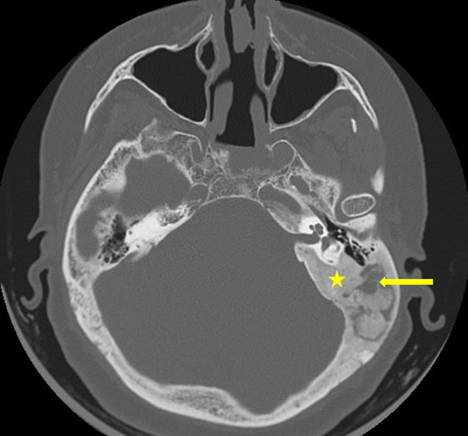
Figure 1. CT scan, bone window and axial view, showing bony overgrowth involving the left temporal bone (yellow star) with sclerosis and lytic changes within the mastoid air cells (yellow arrow).
| Journal of Medical Cases, ISSN 1923-4155 print, 1923-4163 online, Open Access |
| Article copyright, the authors; Journal compilation copyright, J Med Cases and Elmer Press Inc |
| Journal website http://www.journalmc.org |
Case Report
Volume 9, Number 11, November 2018, pages 363-365
Monostotic Fibrous Dysplasia of the Temporal Bone: A Case Report and Review of the Literature
Figures