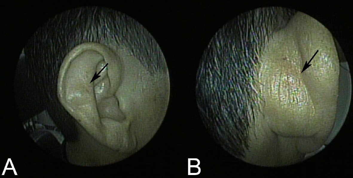
Figure 1. (A) A painless, dome-shaped cystic mass (arrow) in the right auricle. (B) The cystic mass protrudes slightly into the posterior side of auricle.
| Journal of Medical Cases, ISSN 1923-4155 print, 1923-4163 online, Open Access |
| Article copyright, the authors; Journal compilation copyright, J Med Cases and Elmer Press Inc |
| Journal website http://www.journalmc.org |
Case Report
Volume 9, Number 12, December 2018, pages 383-385
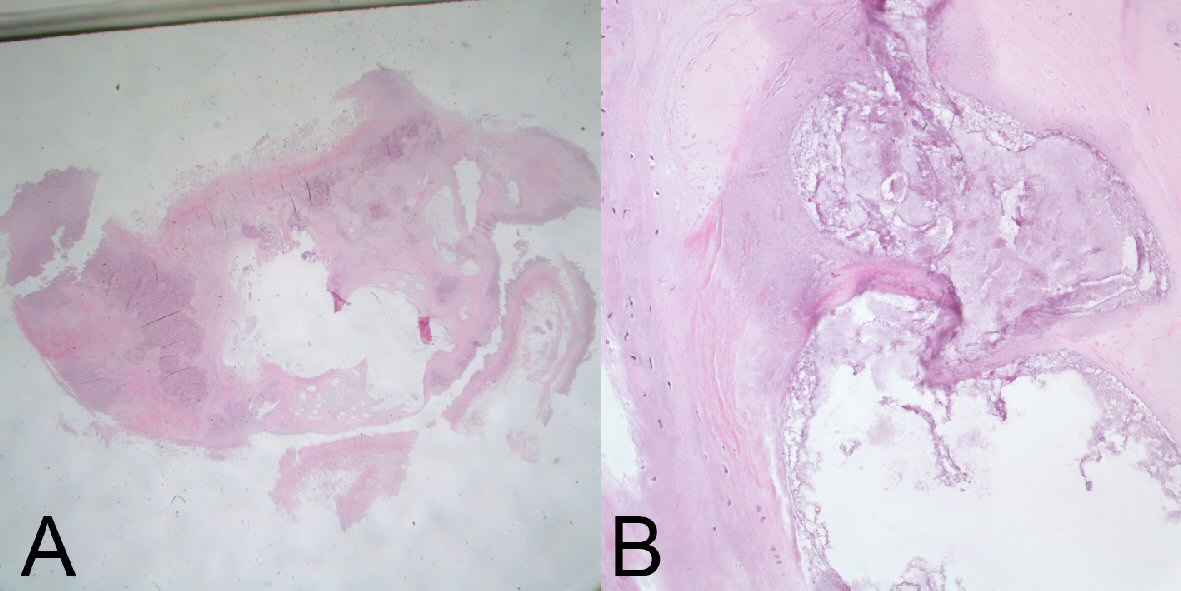
Benign Idiopathic Cystic Chondromalacia Of Auricle
Figures