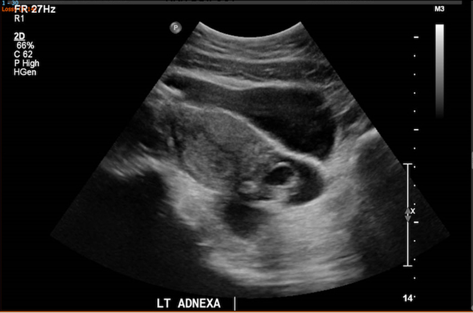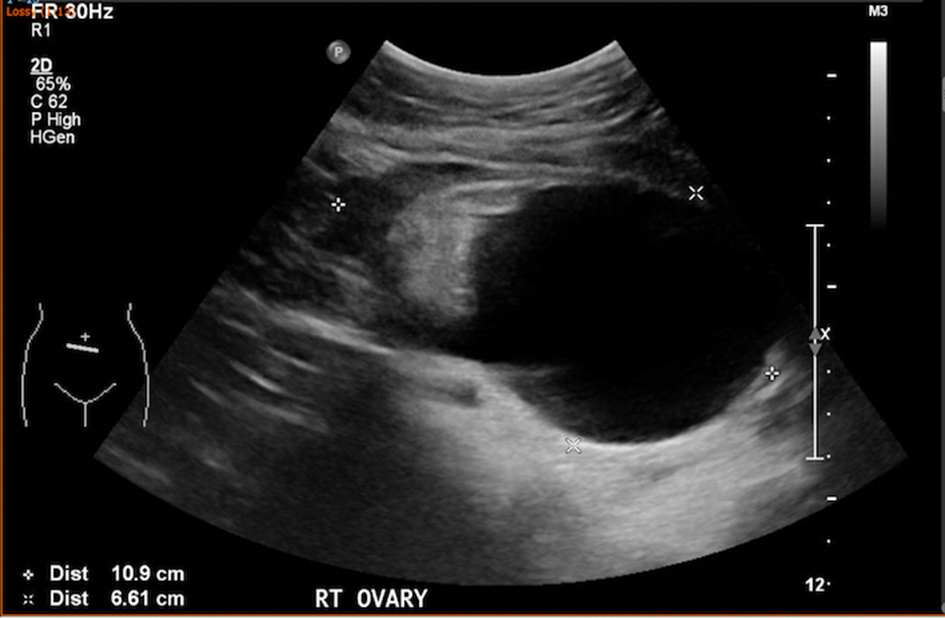
Figure 1. Avascular left adnexal mass of 6.4 × 3.6 × 2.3 cm showed predominantly cystic structure with echogenic components, likely a left ovarian dermoid.
| Journal of Medical Cases, ISSN 1923-4155 print, 1923-4163 online, Open Access |
| Article copyright, the authors; Journal compilation copyright, J Med Cases and Elmer Press Inc |
| Journal website http://www.journalmc.org |
Case Report
Volume 9, Number 12, December 2018, pages 390-393
Laparoscopic Management of a Rare Case of Mature Cystic Teratoma With Immature Neural Tissue and Literature Review
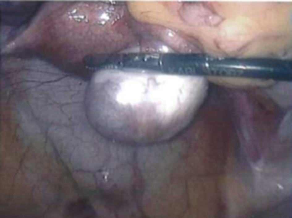
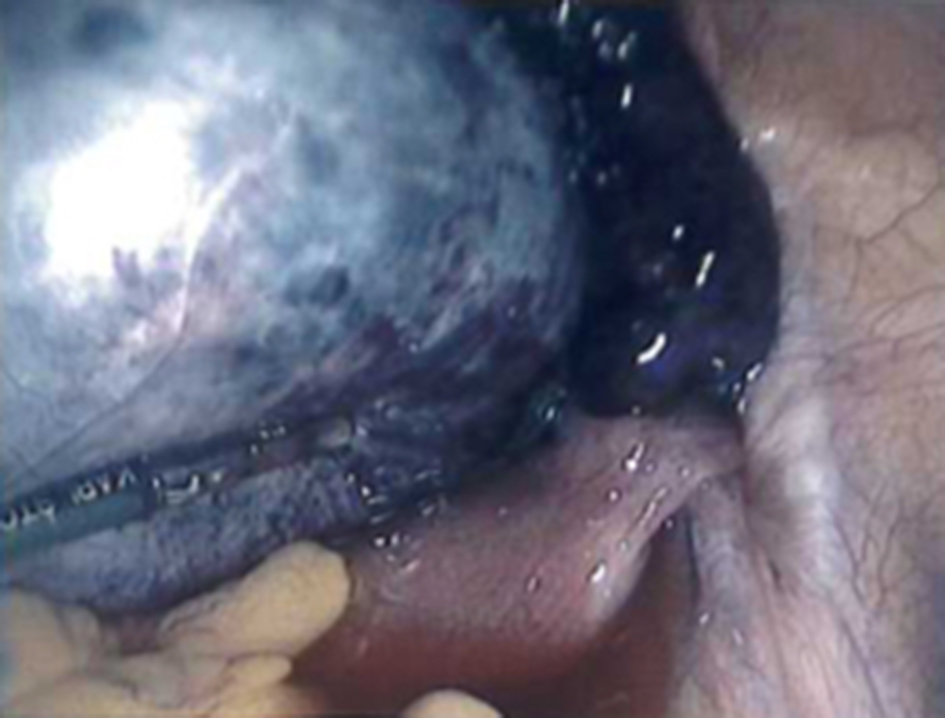
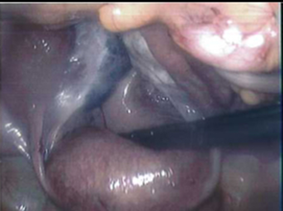
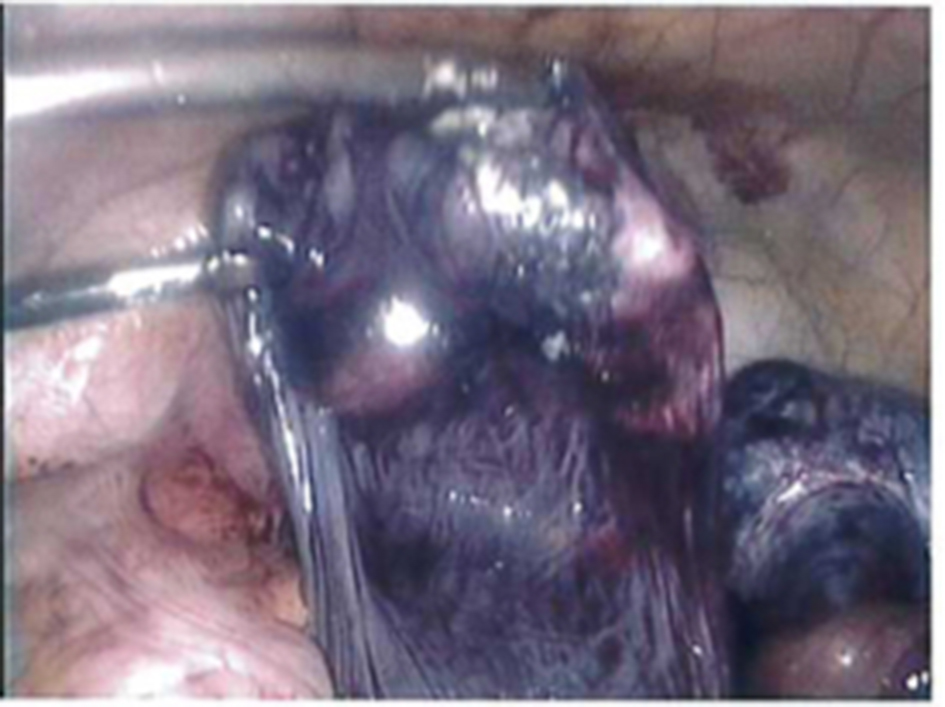
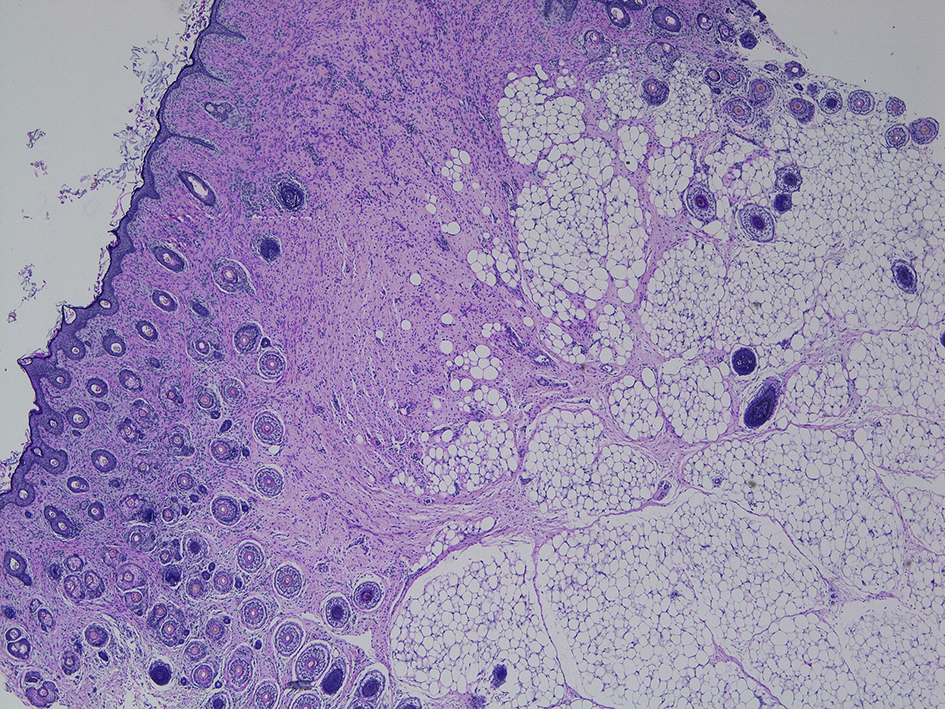
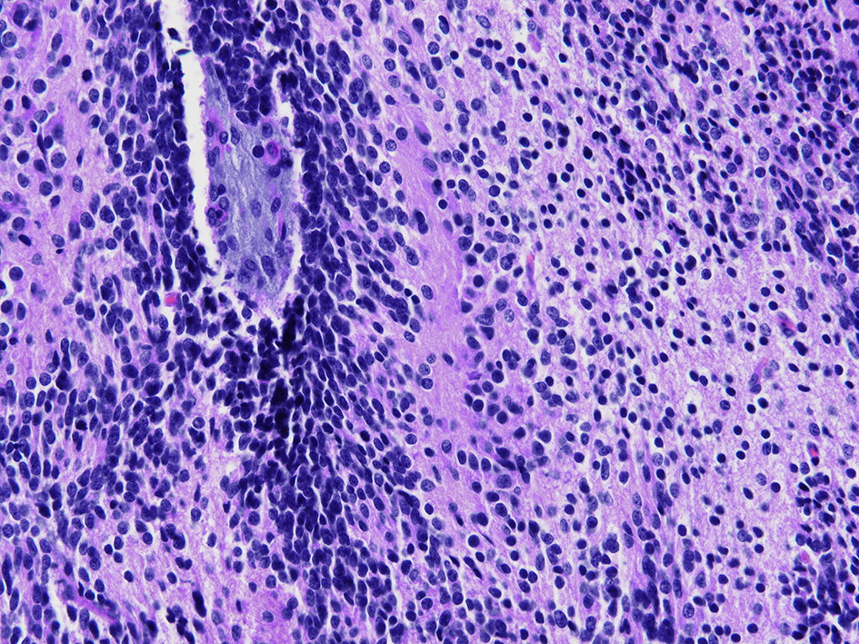
Figures