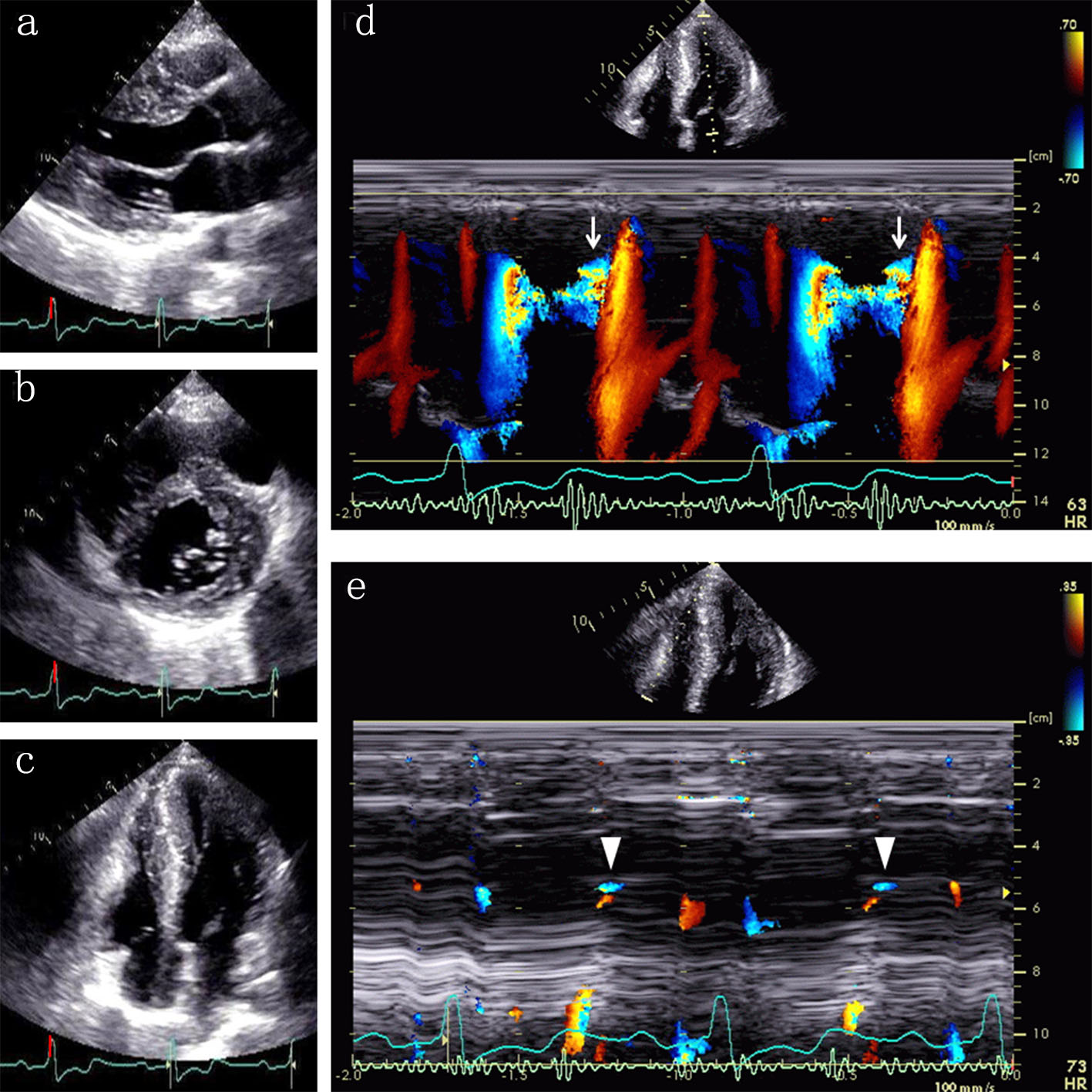
Figure 1. Echocardiographic images at end-diastole show myocardial hypertrophy in the ventricular septum, left ventricular anterior and lateral walls, and the apex of both ventricles ((a) parasternal long axis view; (b) parasternal short axis view; (c) apical four-chamber view). Color Doppler M-mode shows a diastolic paradoxic jet flow from the apex toward the base of the left ventricle ((d) arrows). Note a similar flow in the right ventricle ((e) arrowheads).
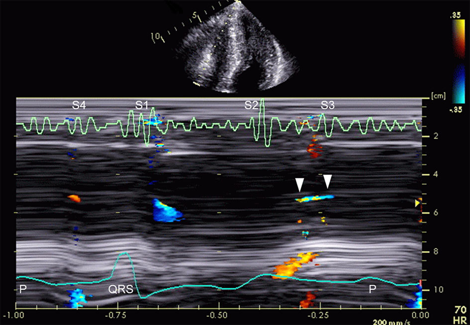
Figure 2. Color Doppler M-mode shows an early diastolic flow from the apex toward the base of the right ventricle (arrowheads). The unique flow lasts to the third heart sound (S3). S1 indicates first heart sound; S2, second heart sound; and S4, fourth heart sound.
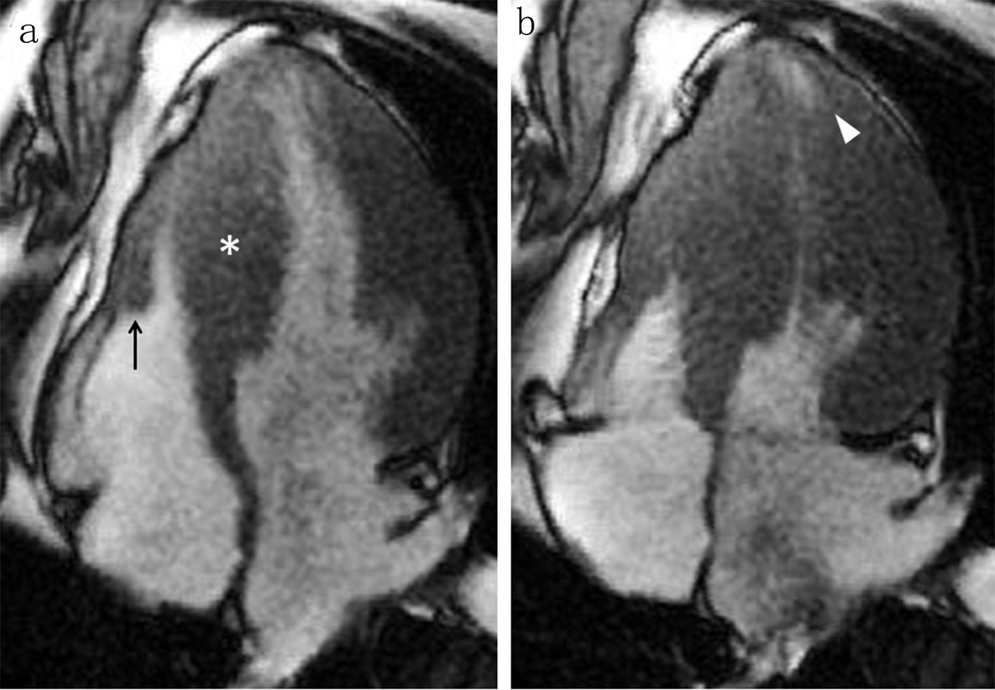
Figure 3. A four-chamber cine cardiac magnetic resonance image at end-diastole shows myocardial hypertrophy in the ventricular septum ((a) asterisk), the right ventricular free wall ((a) arrow), and the left ventricular lateral wall. An apical pouch is present in the left ventricle at end-systole ((b) arrowhead), but not evident in the right ventricle.