Journal of Medical Cases, ISSN 1923-4155 print, 1923-4163 online, Open Access
Article copyright, the authors; Journal compilation copyright, J Med Cases and Elmer Press Inc
Journal website http://www.journalmc.org

Case Report

Volume 10, Number 3, March 2019, pages 67-71


Epidermoid Intramedullary Cyst: A Rare Case Report

Figures

Figure 1.
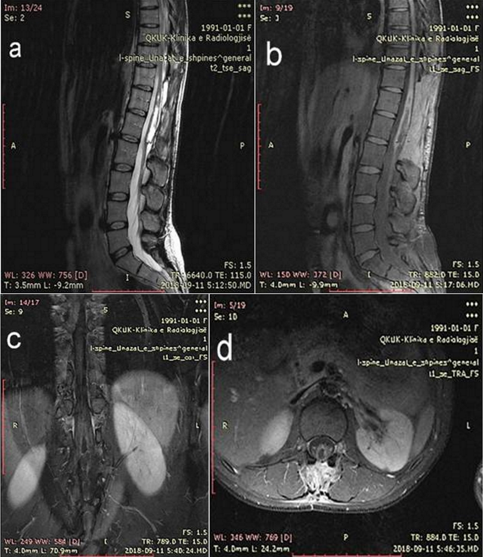
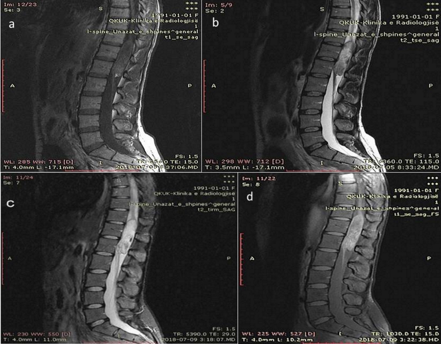
Figure 1. (a) T1-weighted sagital sequence (T11-L1 segments show hypointense, heterogeneous signal). (b) T2-weighted sagital sequence. (c) T2 TIRM sagital sequence. (d) T1/fat sat sagital sequence (T11-L1 segments show hyper intense signal).
Figure 2.
Figure 2. (a) T2-weighted axial sequence (segment T11). (b) T2-weighted axial sequence (segment T12). (c) T2- weighted axial sequence (segment T12/L1).
Figure 3.
Figure 3. (a) T1 fat sat sagital sequence. (b-d) T1 fat sat axial sequence (after administration of intravenous contrast gadoteric acid). The lesion shows minimal peripheral enhancement.
Figure 4.
Figure 4. Histopathology image analysis shows keratin material characteristic for epidermoid cyst.
Figure 5.
Figure 5. (a) T2-weighted sagital sequence. (b) T1- Fat Sat sagital sequence. (c) T1 fat sat coronal sequence. (d)T1 fat sat transversal sequence after administration of intravenous contrast (segments T11-L1). The MRI examination was undertaken after surgical removal of the tumor.