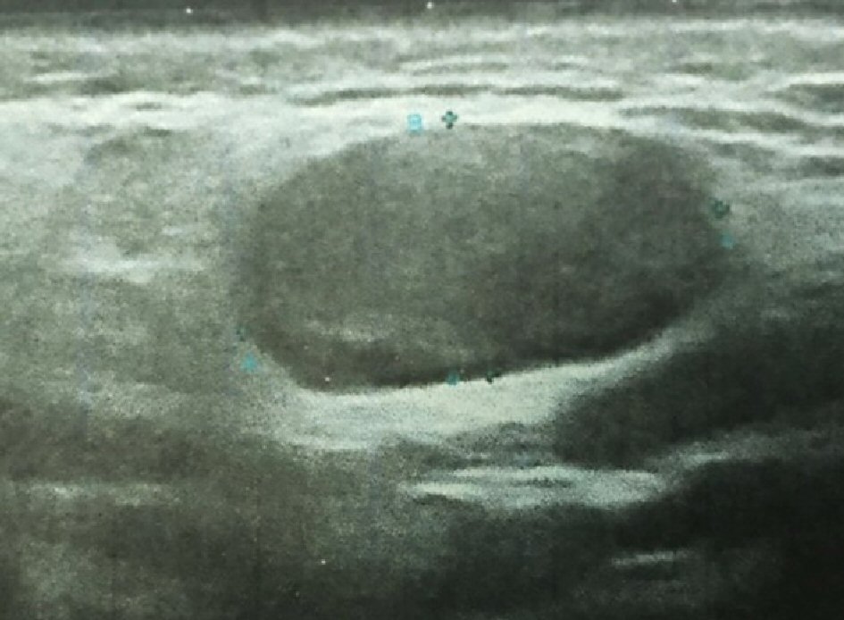
Figure 1. Parotid ultrasonography shows a 20 mm in diameter well-circumscribed mass of the superficial lobe of the right parotid gland. The mass is hypoechoic.
| Journal of Medical Cases, ISSN 1923-4155 print, 1923-4163 online, Open Access |
| Article copyright, the authors; Journal compilation copyright, J Med Cases and Elmer Press Inc |
| Journal website http://www.journalmc.org |
Case Report
Volume 10, Number 5, May 2019, pages 146-149
Parotid Gland Oncocytoma: A Rare Case and Literature Review
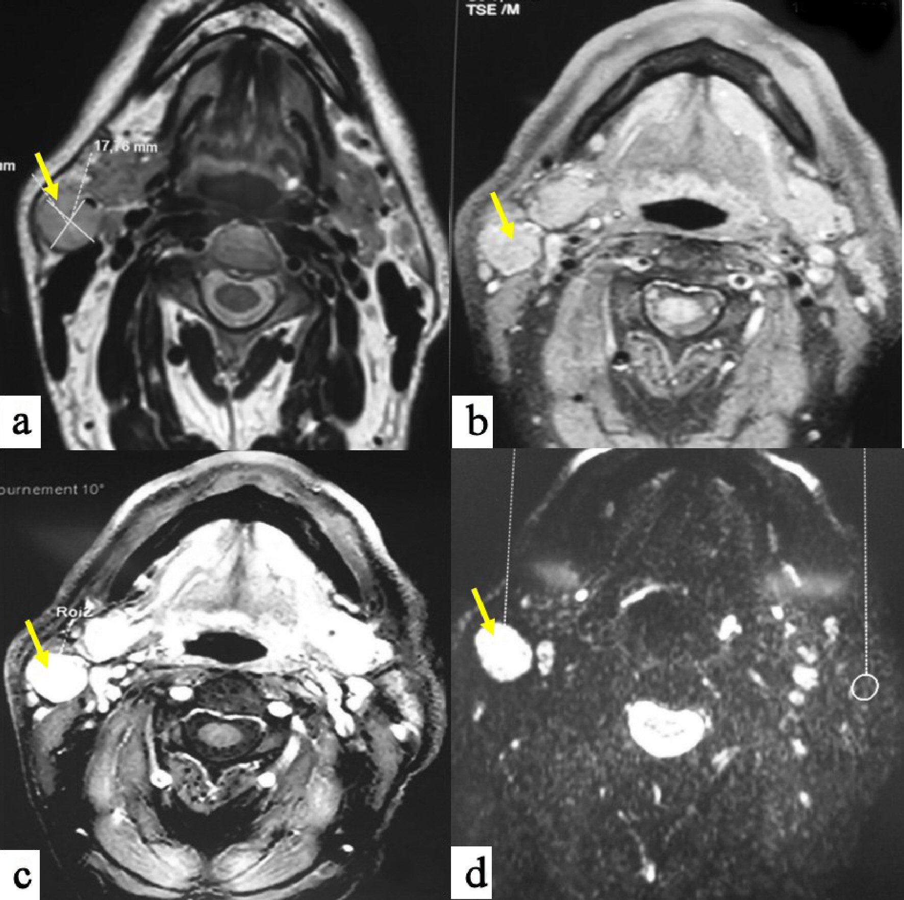
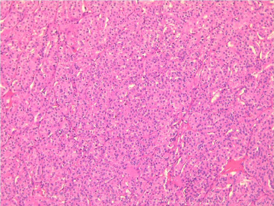
Figures