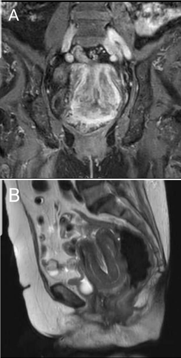
Figure 1. Coronal (A) and sagittal (B) MRI views showing U-shaped uterine cavity with thickened and inverted uterine fundus.
| Journal of Medical Cases, ISSN 1923-4155 print, 1923-4163 online, Open Access |
| Article copyright, the authors; Journal compilation copyright, J Med Cases and Elmer Press Inc |
| Journal website http://www.journalmc.org |
Case Report
Volume 10, Number 4, April 2019, pages 110-112
Idiopathic Postmenopausal Uterine Inversion: A Case Report
Figures