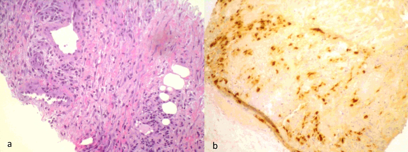
Figure 1. Mediastinal lesion biopsy: areas with vascular structures with perivascular epithelioid-like cells (a), which showed positivity for the HMB45 in the immunohistochemical study compatible with LAM cells (b).
| Journal of Medical Cases, ISSN 1923-4155 print, 1923-4163 online, Open Access |
| Article copyright, the authors; Journal compilation copyright, J Med Cases and Elmer Press Inc |
| Journal website http://www.journalmc.org |
Case Report
Volume 10, Number 7, July 2019, pages 218-221
Lymphangioleiomyomatosis: A Challenging Case
Figures
Table
| FEV 1 (%) | TI (%) | DLCO (%) | SMWT/SpO2 (m/%) | PaO2 at rest (mm Hg) | Supplementary O2 at rest/deambulation | |
|---|---|---|---|---|---|---|
| FEV1: forced expiratory volume in 1 s; TI: Tiffeneau index; DLCO: diffusing capacity of the lungs for carbon monoxide; SMWT: six-minute walk test; SpO2: peripheral capillary oxygen saturation; PaO2: partial pressure of arterial oxygen. aStarted treatment with sirolimus. | ||||||
| 01-2014 | 40.4 | 75.07 | 45.1 | -/89% | 70 | No/No |
| 10-2016 | 44.3 | 65.07 | 43.9 | 460/82% | 64 | No/No |
| 01-2017 | 40.8 | 63.2 | 44 | - | - | No/Yes |
| 07-2017a | 38.7 | 66.7 | 33.8 | - | 53 | Yes/Yes |
| 11-2017 | 49.1 | 69.3 | 52 | -/86% | 84 | No/Yes |
| 01-2018 | 45.7 | 62.55 | 43 | - | 65 | No/Yes |
| 03-2018 | 61 | 76.54 | 50.3 | - | 69 | No/Yes |
| 06-2018 | 52 | 68.77 | 46 | 420 m/86% | 83 | No/Yes |
| 09-2018 | 60 | 80.3 | 45 | - | 72 | No/Yes |
| 12-2018 | 66 | 78.36 | 49 | - | - | No/Yes |
| 03-2019 | 60 | 74.14 | 44 | - | 78 | No/Yes |