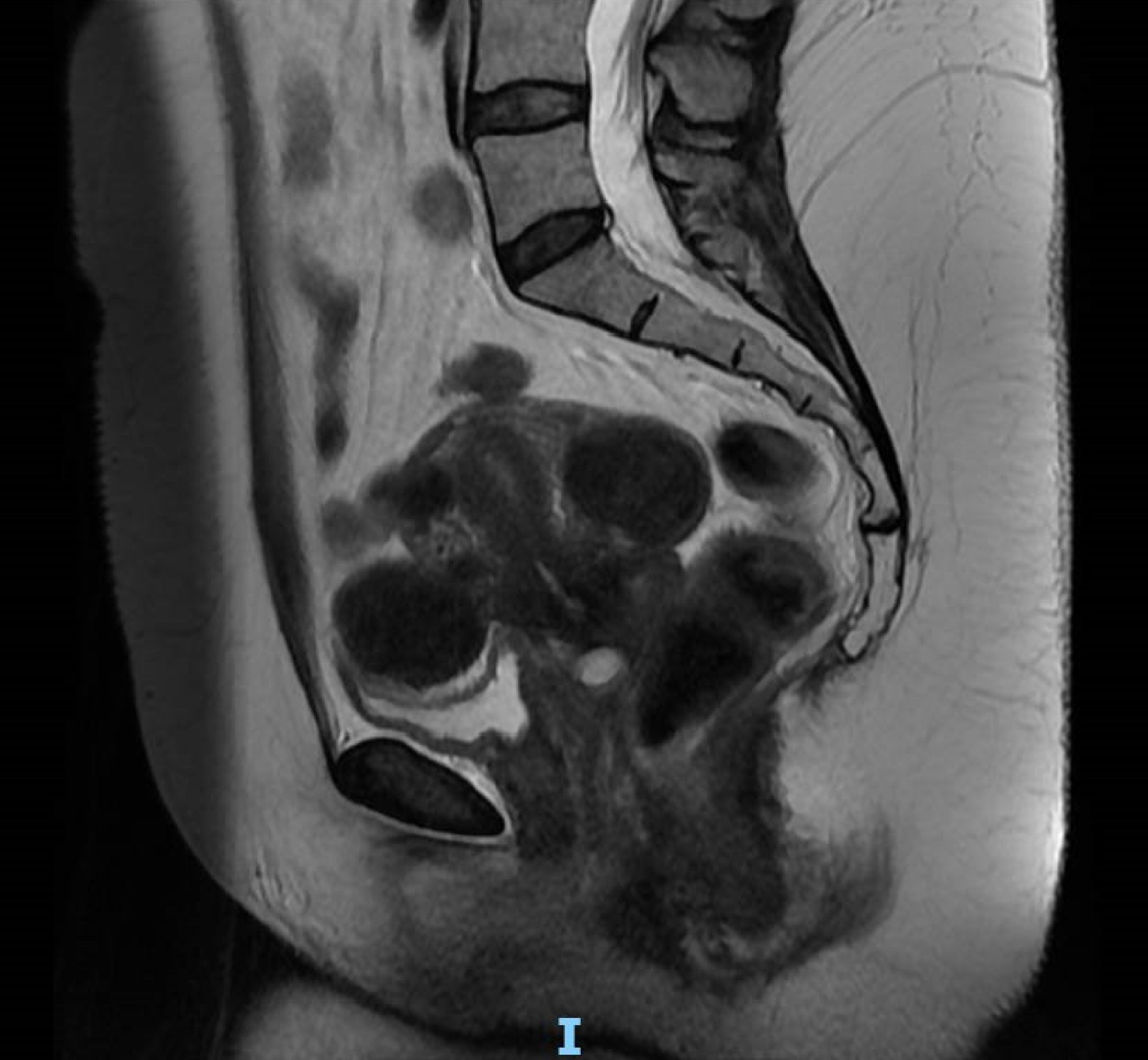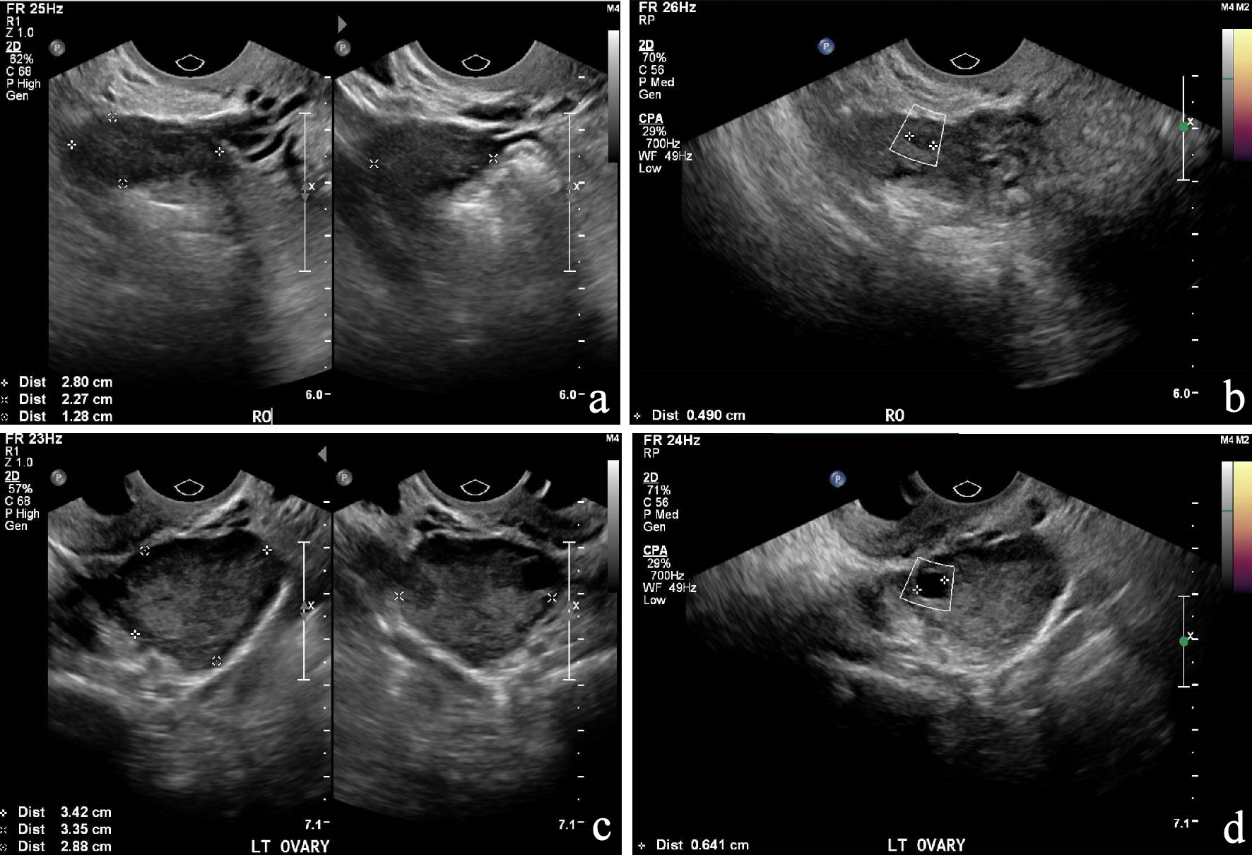
Figure 1. (a) and (b): US of pelvis of the right ovary; (c) and (d): US of pelvis of the left ovary. US: ultrasonography.
| Journal of Medical Cases, ISSN 1923-4155 print, 1923-4163 online, Open Access |
| Article copyright, the authors; Journal compilation copyright, J Med Cases and Elmer Press Inc |
| Journal website http://www.journalmc.org |
Case Report
Volume 10, Number 11, November 2019, pages 323-327
A Rare Case of Ovarian Follicular Lymphoma: Incidental Finding in a Woman With Postmenopausal Bleeding
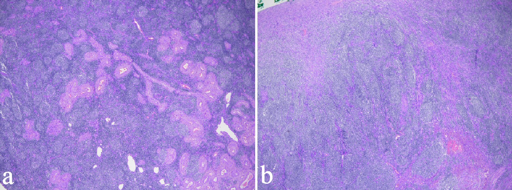
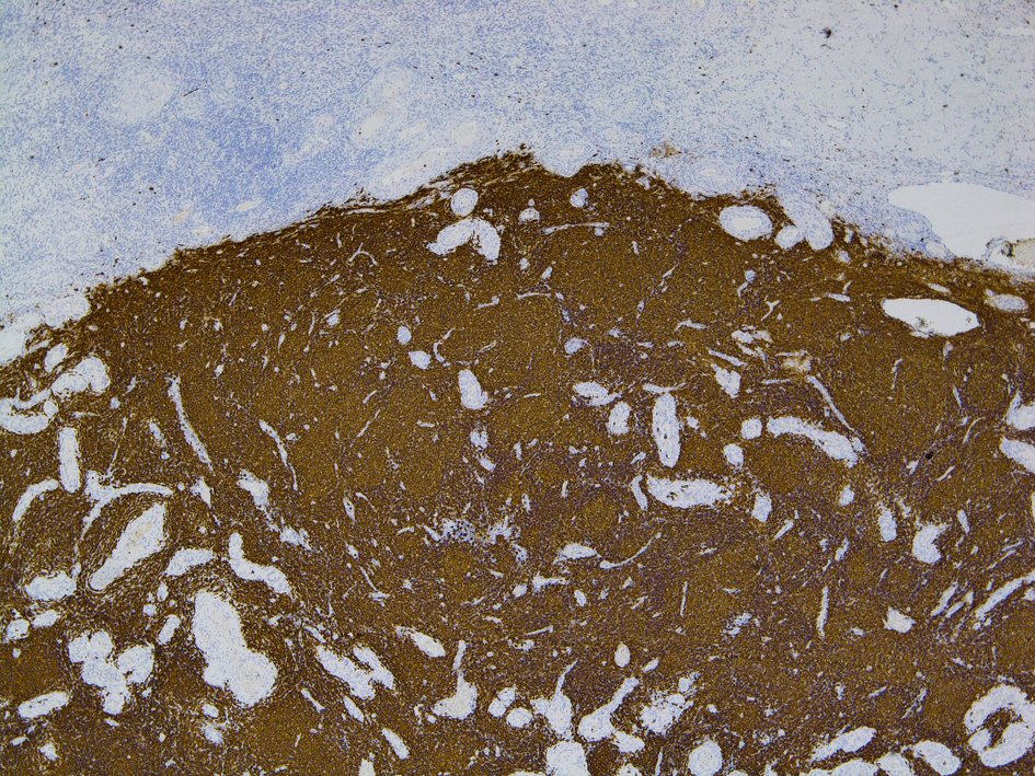
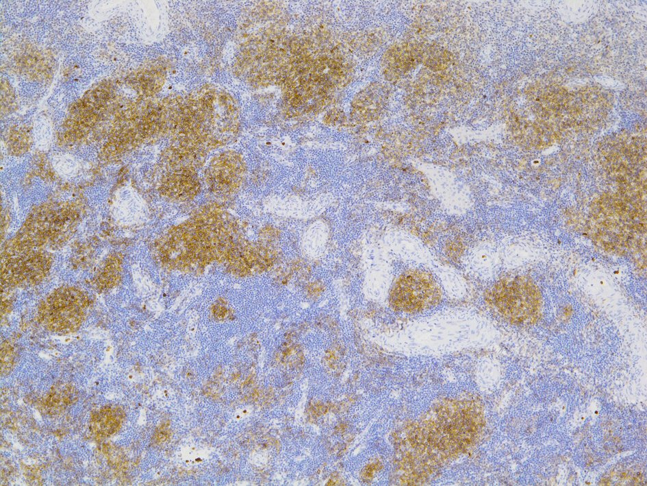
Figures