Journal of Medical Cases, ISSN 1923-4155 print, 1923-4163 online, Open Access
Article copyright, the authors; Journal compilation copyright, J Med Cases and Elmer Press Inc
Journal website http://www.journalmc.org

Case Report

Volume 11, Number 3, March 2020, pages 73-76


Carcinoid Right Heart Disease

Figures

Figure 1.
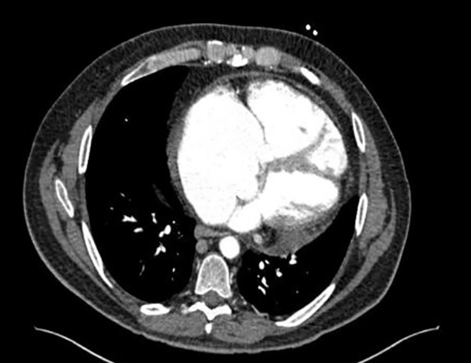
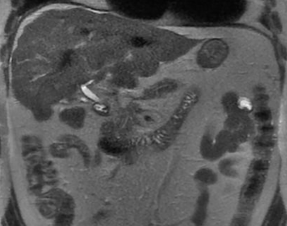
Figure 1. Cirrhotic liver morphology. Multiple low-attenuation foci in the liver (likely treated metastases). Necrotic foci in the peripancreatic space.
Figure 2.
Figure 2. Severely dilated right atrium and ventricle.
Figure 3.
Figure 3. Right ventricle (RV) severely dilated. RV systolic tissue Doppler velocity is 9.0 cm/s. The estimated right ventricular systolic pressure is 43 mm Hg consistent with mild pulmonary hypertension (but may be underestimated due to severe tricuspid regurgitation).
Figure 4.
Figure 4. Severe (4+) tricuspid valve regurgitation.
Figure 5.
Figure 5. Status post valves replacement. Biocor prosthetic tricuspid valve size #33. Biocor prosthetic pulmonary valve size #27. There is no prosthetic regurgitation.