Journal of Medical Cases, ISSN 1923-4155 print, 1923-4163 online, Open Access
Article copyright, the authors; Journal compilation copyright, J Med Cases and Elmer Press Inc
Journal website http://www.journalmc.org

Case Report

Volume 11, Number 5, May 2020, pages 111-114


A Neglected Case of Colonic Tuberculosis With Thoracolumbar Enterocutaneous Fecal Fistulae

Figures

Figure 1.
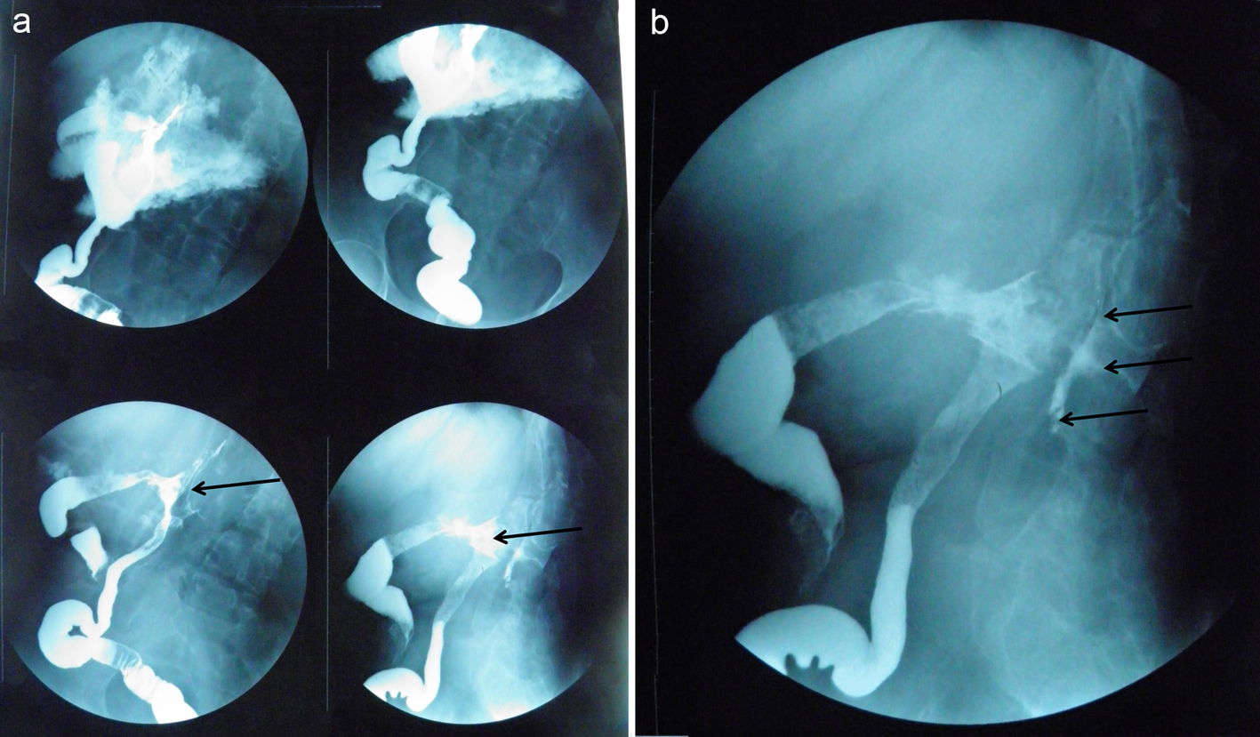
Figure 1. (a) Digestive tract barium studies showing fistulae (black arrows) between the colon and the thoracolumbar structures. (b) Fistulography showing multiple fistulae (black arrows) connecting the bowel lumen of the proximal descending colon to the exterior of the posterior abdominal wall.
Figure 2.
Figure 2. Colonic mucosa at colonoscopy: the fecal fistula aperture is clearly identified.
Figure 3.
Figure 3. Photomicrograph of the intraoperative histopathology showing typical granulomatous features of tuberculosis: M: multinucleate giant cells; C: area of caseation within a granuloma; G: granuloma; L: lymphocytic infiltrate.