Journal of Medical Cases, ISSN 1923-4155 print, 1923-4163 online, Open Access
Article copyright, the authors; Journal compilation copyright, J Med Cases and Elmer Press Inc
Journal website http://www.journalmc.org

Case Report

Volume 11, Number 5, May 2020, pages 125-128


Atropine-Induced Ventricular Asystole During a Dobutamine Stress Test

Figures

Figure 1.
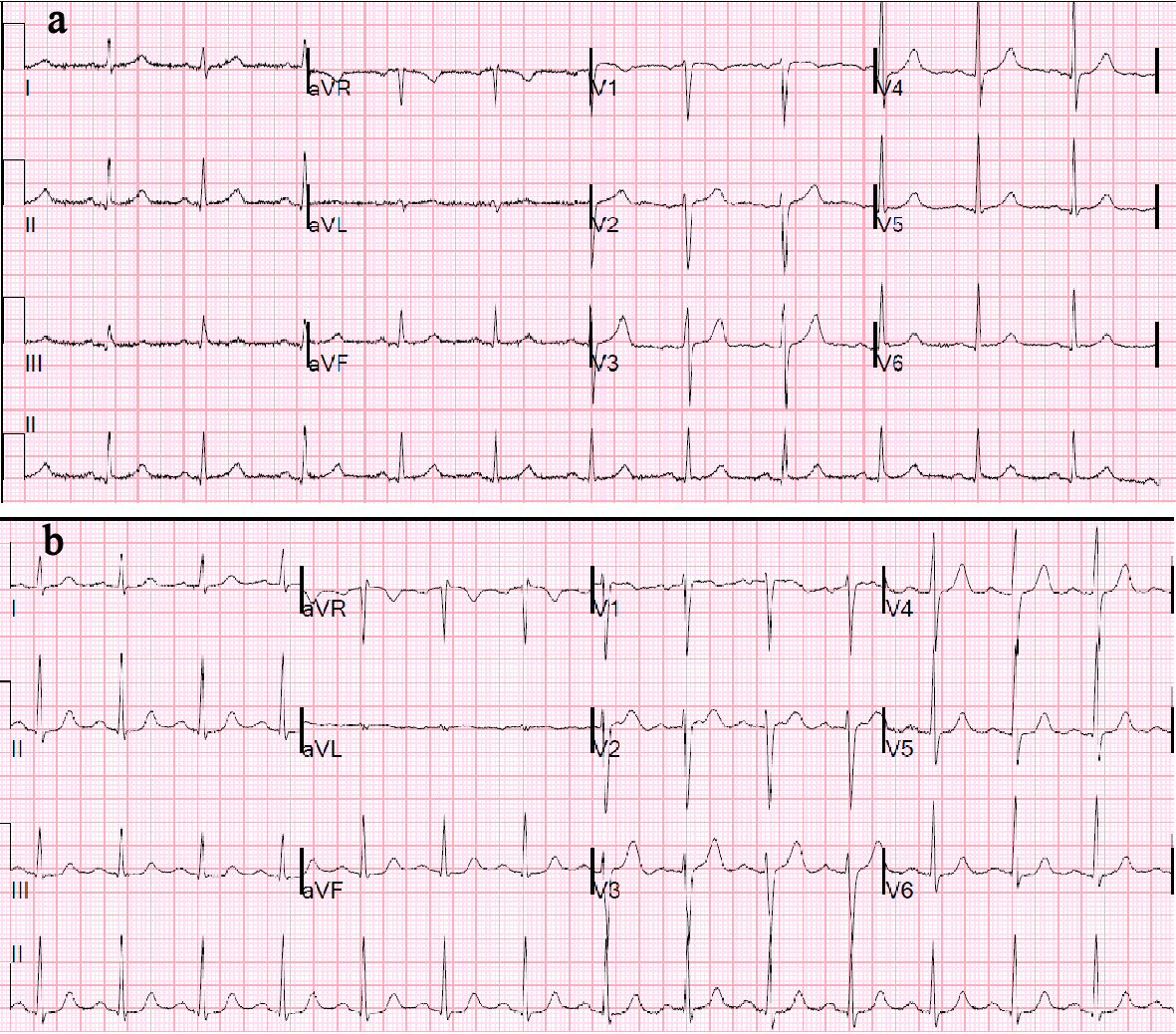
Figure 1. (a) Baseline ECG with NSR, LVH, and LAA, PR = 0.19 s. (b) ECG after second episode of third-degree AV block (PR = 0.22 s), which demonstrates new first-degree AVB. ECG: electrocardiogram; AVB: atrioventricular block; LVH: left ventricular hypertrophy; NSR: normal sinus rhythm.
Figure 2.
Figure 2. (a) ECG during DSE showing prolonging PR interval followed by complete heart block and ventricular asystole due to lack of escape rhythm. (b) Telemetry strip post-admission showing the same rhythm. ECG: electrocardiogram; DSE: dobutamine stress echocardiography.