Journal of Medical Cases, ISSN 1923-4155 print, 1923-4163 online, Open Access
Article copyright, the authors; Journal compilation copyright, J Med Cases and Elmer Press Inc
Journal website http://www.journalmc.org

Case Report

Volume 11, Number 6, June 2020, pages 178-181


Percutaneous Endoscopic Surgery for Lumbar Discal Cyst: Two Case Reports

Figures

Figure 1.
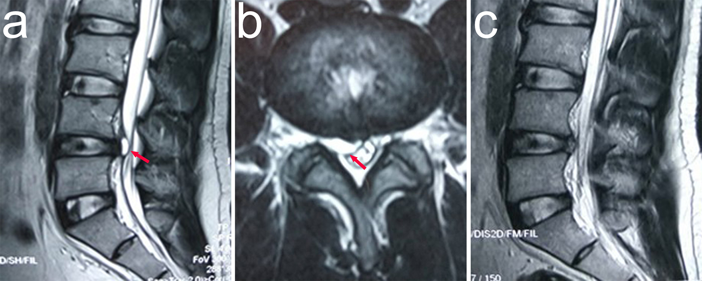
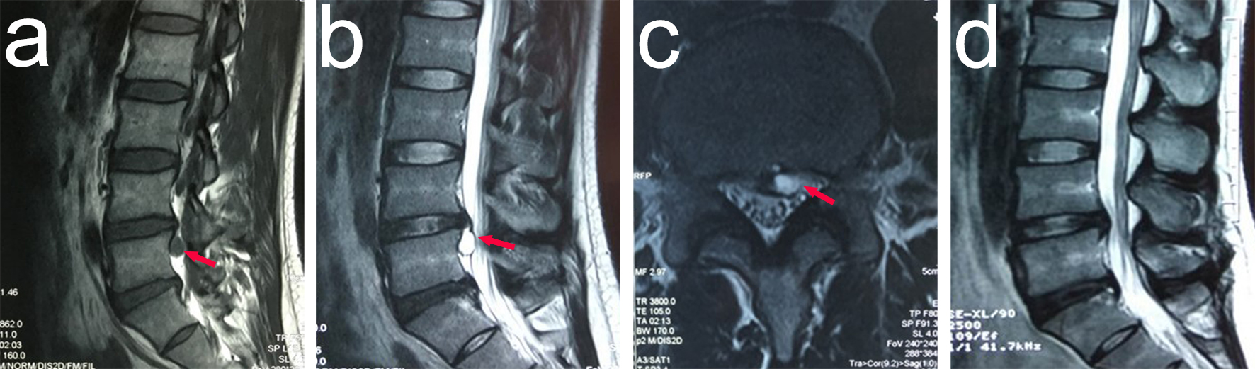
Figure 1. Lumbar spinal MRI of the patient demonstrating the L4-L5 discal cyst, with prominent compression of the left L5 nerve root. (a) Sagittal T1-weighted image shows an ovoid lesion with low signal intensity (red arrow). (b) Sagittal T2-weighted image shows an ovoid lesion with a hyperintense center plus hypointense rim (red arrow). (c) Axial MRI shows the lesion (red arrow). (d) Lumbar T2-weighted MRI shows no residual cyst. MRI: magnetic resonance imaging.
Figure 2.
Figure 2. Lumbar spinal MRI of the patient demonstrating the L4-L5 discal cyst, with prominent compression of the right L5 nerve root. (a) Sagittal T2-weighted image shows an ovoid lesion with a hyperintense center plus hypointense rim (red arrow). (b) Axial MRI shows the lesion (red arrow). (c) Lumbar T2-weighted MRI shows no residual cyst. MRI: magnetic resonance imaging.