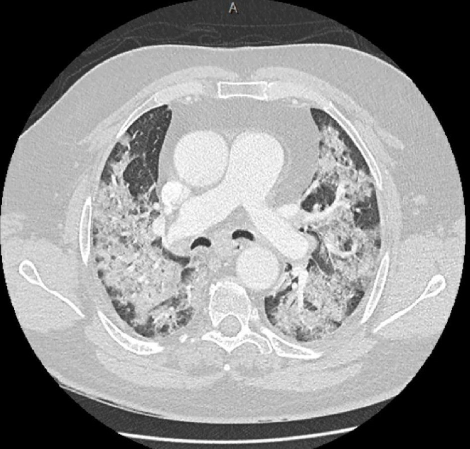
Figure 1. Computed Tomography angiogram of the thorax showing extensive bilateral patchy and ground glass opacities throughout the lungs without any pulmonary embolism.
| Journal of Medical Cases, ISSN 1923-4155 print, 1923-4163 online, Open Access |
| Article copyright, the authors; Journal compilation copyright, J Med Cases and Elmer Press Inc |
| Journal website http://www.journalmc.org |
Case Report
Volume 11, Number 7, July 2020, pages 189-191
A Case of COVID-19 Complicated by Venous Thromboembolism Despite Being on Prophylactic Anticoagulation
Figures