Journal of Medical Cases, ISSN 1923-4155 print, 1923-4163 online, Open Access
Article copyright, the authors; Journal compilation copyright, J Med Cases and Elmer Press Inc
Journal website http://www.journalmc.org

Case Report

Volume 11, Number 8, August 2020, pages 234-238


Cor Triatriatum Dexter: A Case Report in a 70-Year-Old Male

Figures

Figure 1.
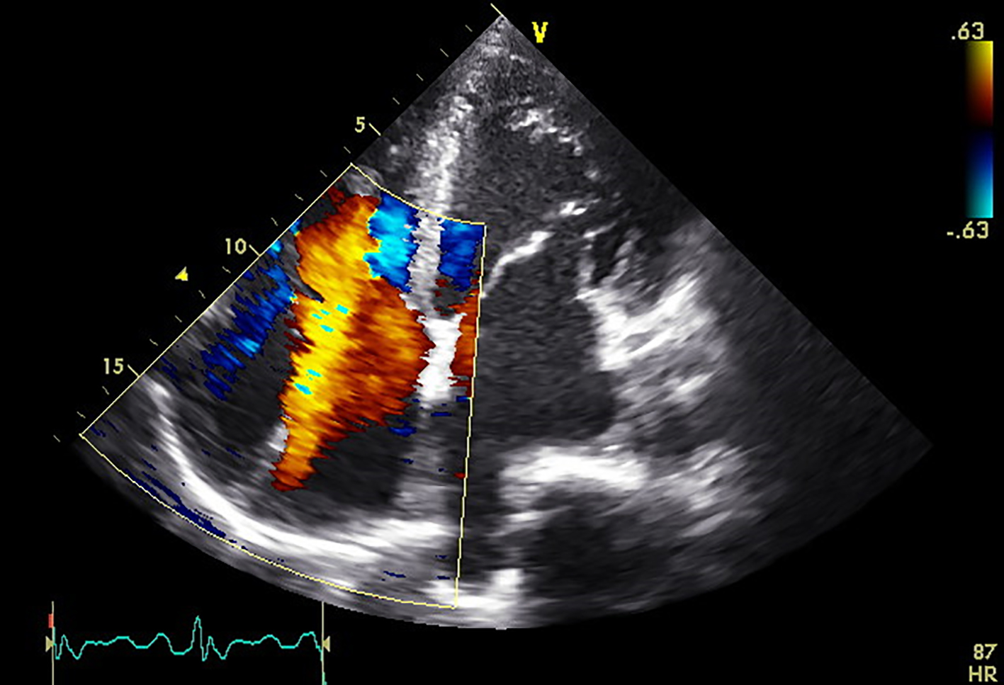
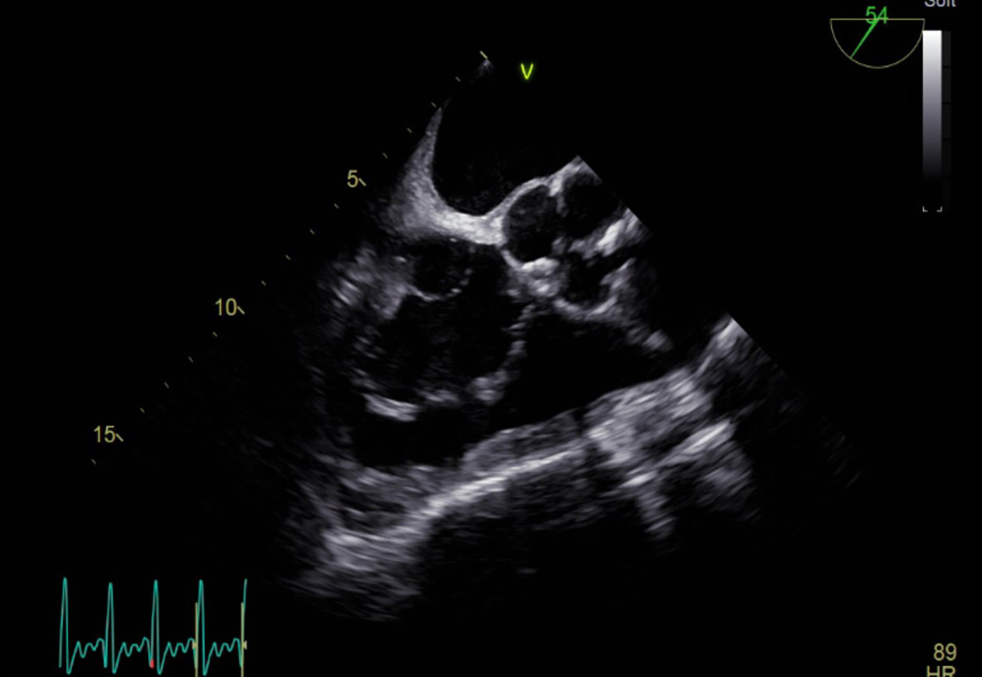
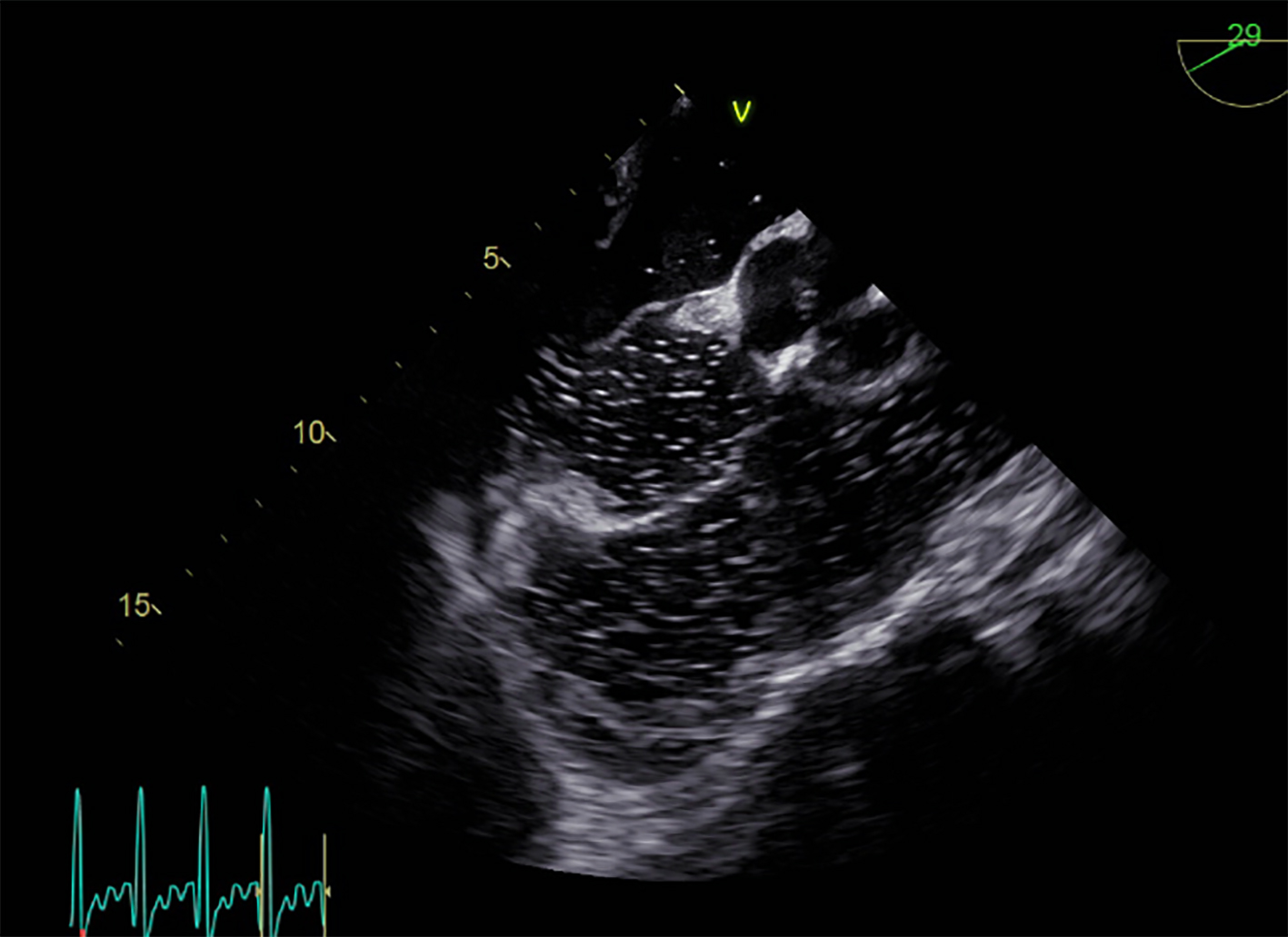
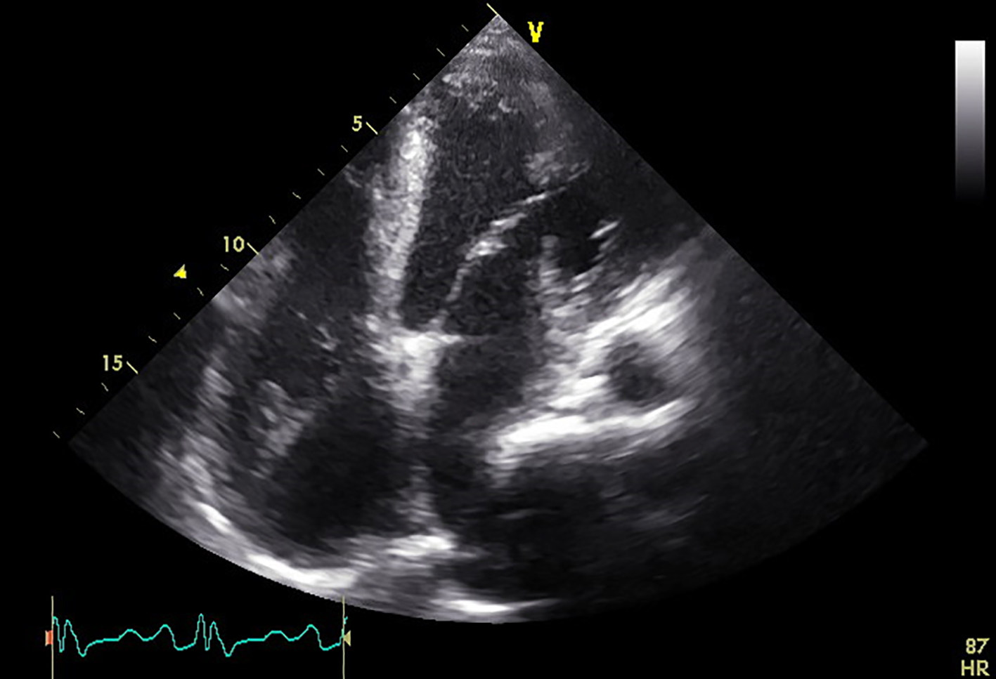
Figure 1. Transthoracic echocardiography in an apical four-chamber view revealing a left ventricular ejection fraction of 65% and a possible septation abnormality within the right atrium.
Figure 2.
Figure 2. Transthoracic echocardiography in an apical four-chamber view with color Doppler revealing moderate to severe tricuspid regurgitation.
Figure 3.
Figure 3. Transesophageal echocardiography at the level of the aortic valve with the scope rotated to 54° showing evidence of a septum within the right atrium diagnosed as cor triatriatum dexter.
Figure 4.
Figure 4. Transesophageal echocardiography at the level of the aortic valve with the scope rotated to 29° during a bubble study, revealing an intra-atrial septal aneurysm with a small patent foramen ovale and a right-to-left shunt as evidenced by the bubbles within the left atrium.