Journal of Medical Cases, ISSN 1923-4155 print, 1923-4163 online, Open Access
Article copyright, the authors; Journal compilation copyright, J Med Cases and Elmer Press Inc
Journal website https://www.journalmc.org

Case Report

Volume 12, Number 4, April 2021, pages 149-151


Giant Cell Tumor of the Temporal Bone With Skull Base and Middle Ear Extension

Figures

Figure 1.
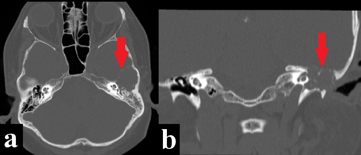
Figure 1. Temporal bone computed tomography (CT) bone window shows a destructive and expansive process eroding the left middle skull base, tegmen tympani, ossicular chain, Scutum, basal turn of the cochlea, and tympanic portion of the facial nerve (red arrows). (a) Axial view. (b) Coronal view.
Figure 2.
Figure 2. Magnetic resonance imaging (MRI). (a) Axial T2-FLAIR sequence shows solid extra-axial mass with low signal (white arrow). (b, c) Axial and coronal T1 Fast Sat sequence showing moderate enhancement after intravenous contrast administration (yellow arrows).
Figure 3.
Figure 3. H&E stain: fragments of stromal lesion with osteoclast- like giants cells. H&E: hematoxylin and eosin.