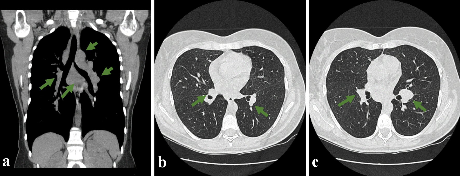
Figure 1. Thoracic CT scan showing multiple mediastinal adenopathies, in all compartments, with the largest ones standing out subcarinal (a), about 37 mm in diameter, and a 32 mm pre-tracheal in diameter. (a) Coronal section showing through the green arrows several mediastinal adenopathies; (b, c) Cross-section to show in the pulmonary window the presence of multiple adenopathies and pulmonary nodules (green arrow). CT: computed tomography.