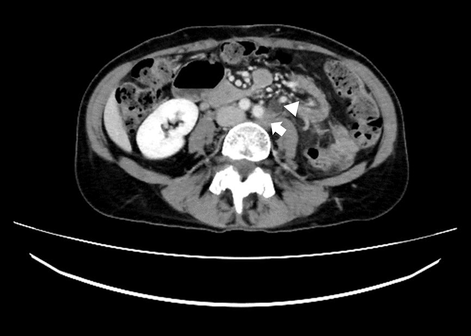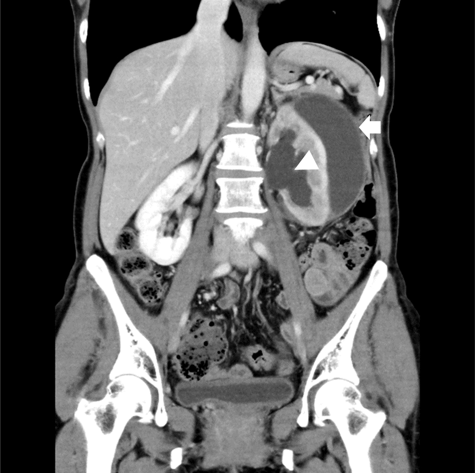
Figure 1. A coronal view on CECT scan revealed a low attenuation fluid collection in the subcapsular area (arrow) in the setting of dilation of the renal pelvis (arrowhead). CECT: contrast-enhanced computed tomography.
| Journal of Medical Cases, ISSN 1923-4155 print, 1923-4163 online, Open Access |
| Article copyright, the authors; Journal compilation copyright, J Med Cases and Elmer Press Inc |
| Journal website https://www.journalmc.org |
Case Report
Volume 12, Number 3, March 2021, pages 126-129
Renal Subcapsular Hematoma Formation Due to Hydronephrosis Caused by Recurrent Uterine Cervical Cancer
Figures