| Ikari (2010, Japan) [11] | 67, M | Irregular fibrosis, lymphadenopathy | Lobectomy | Castleman’s disease | 2,280 | ND | SQ (pT1N0M0) (lobectomy) | Dead (> 15 months) |
| Inoue (2014, Japan) [12] | 78, M | GGO, consolidation, lymphadenopathy | Lobectomy and partial resection (for biopsy) | Gastric cancer, autoimmune pancreatitis | 983 | ND | Ad (pT1bN0M0) (lobectomy) | No recurrence (18 months) |
| Tashiro (2016, Japan) [14] | 72, M | A small nodule | Lobectomy | Storiform fibrosis, retroperitoneal fibrosis | 346 | > 40% | Ad (cT1bN0M0) (lobectomy) | No recurrence (5 years) |
| Gomez-Hernandez (2018, Spain) [10] | 70, F | A mass with mediastinal lymphadenopathy | Lobectomy | Pericarditis | ND | ND | Ad (pT1aN2M0) (lobectomy) | ND |
| Tokuda (2018, Japan) [16] | 71, M | Bilateral lung nodules, hilar and mediastinal lymphadenopathy | Bronchoscopy | None | ND | ND | Ad (cT3N3M1b) (chemotherapy) | Dead (1 year and 5 months) |
| Abbass (2019, USA) [8] | 64, M | Multiple pulmonary nodules | CTGB, VATS | PR3-ANCA(+)GPA | 153 | > 10% | SCLC (ND) (chemotherapy) | Dead (10 months) |
| Choi (2019, South Korea) [9] | 66, M | A subpleural nodule in reticular and honeycomb fibrosis | Lobectomy | Autoimmune cholangitis, IPF | 232.4 | > 40% | AdSq (pT2aN2M0) (lobectomy) | ND |
| Terashima (2020, Japan) [15] | 64, M | Bilateral pleural effusion | Pleural biopsy | Eosinophilia, serum IgE↑ | 2,750 | > 50% | Ad (stage IIIB) (CRT → durvalumab) | ND |
| Ito (2020, Japan) [13] | 80, M | Systemic lymphadenopathy | Axillary lymph node biopsy | None | 137 | > 40% | Ad (cT3N3M0) (CRT → osimertinib) | ND |
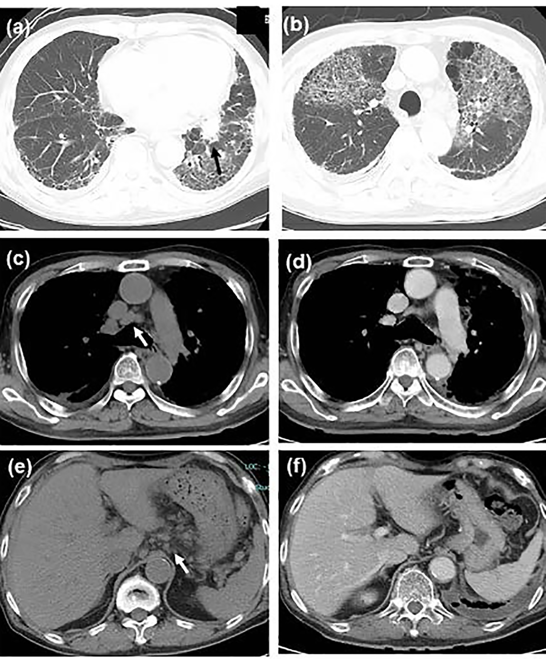
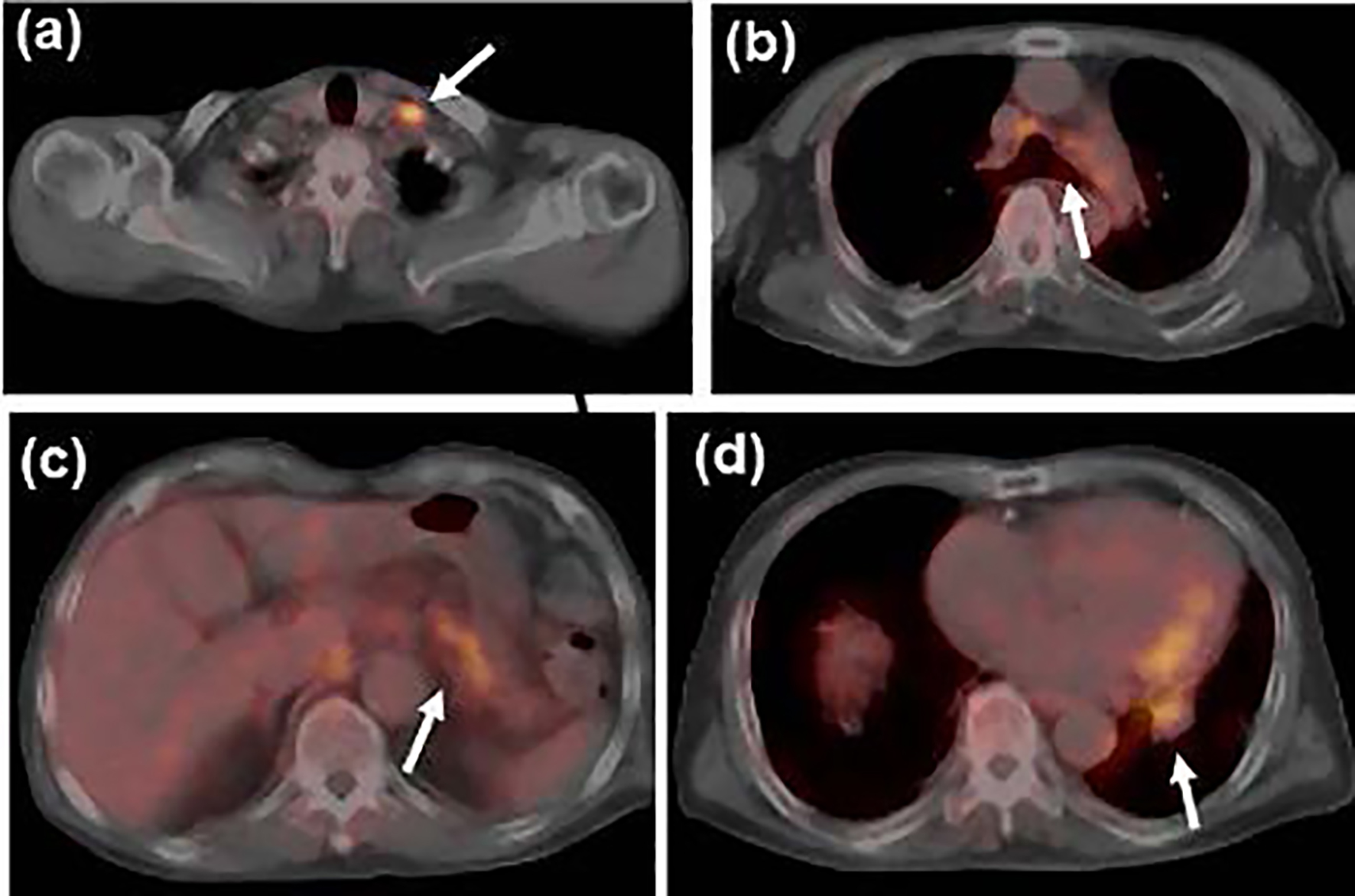
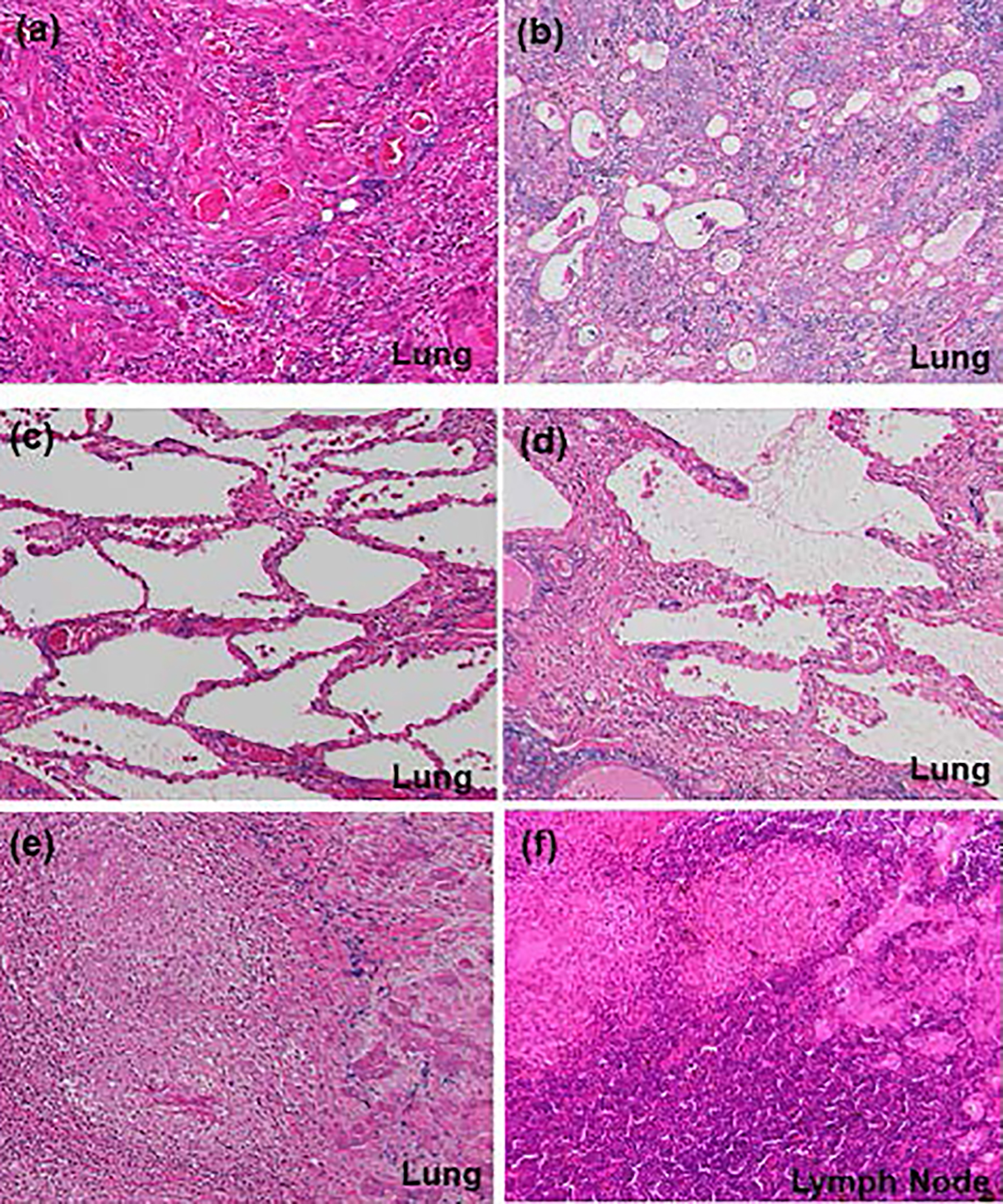
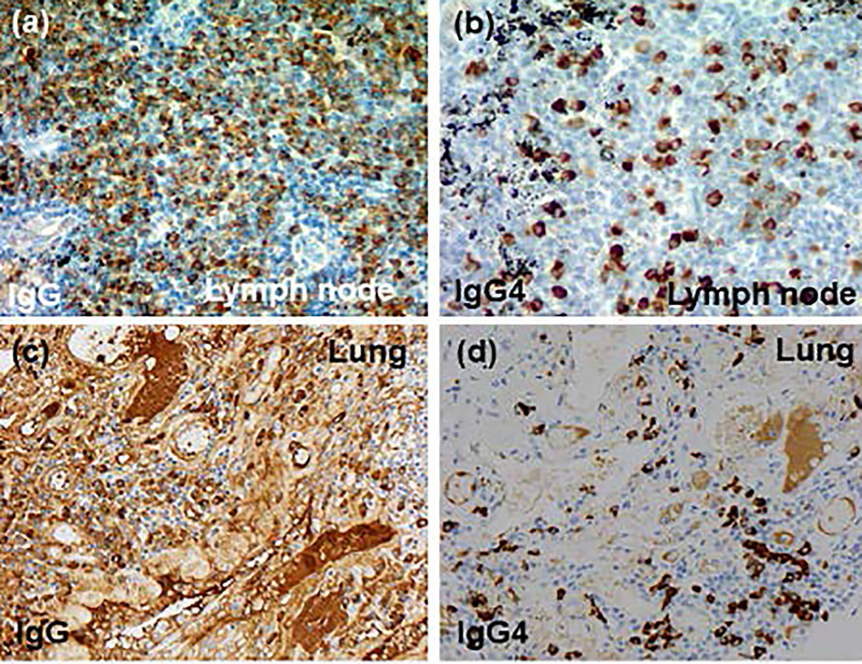
| Ours (Japan) | 75, M | A tumor, interstitial pneumonia, and systemic lymphadenopathy | Lobectomy | NSIP, tuberculosis, pneumothorax | 385 | 54% | AdSq (pT2aN2M0) (lobectomy) | Dead (24 days) |