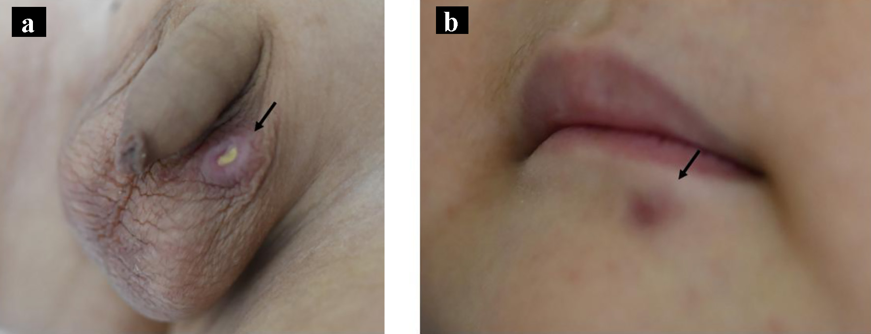
Figure 1. Macroscopic appearance of the tumors on the left scrotum (a, arrow) and under the lip (b, arrow).
| Journal of Medical Cases, ISSN 1923-4155 print, 1923-4163 online, Open Access |
| Article copyright, the authors; Journal compilation copyright, J Med Cases and Elmer Press Inc |
| Journal website https://www.journalmc.org |
Case Report
Volume 12, Number 8, August 2021, pages 306-309
Lymphomatoid Papulosis Development in Acute Lymphoblastic Leukemia
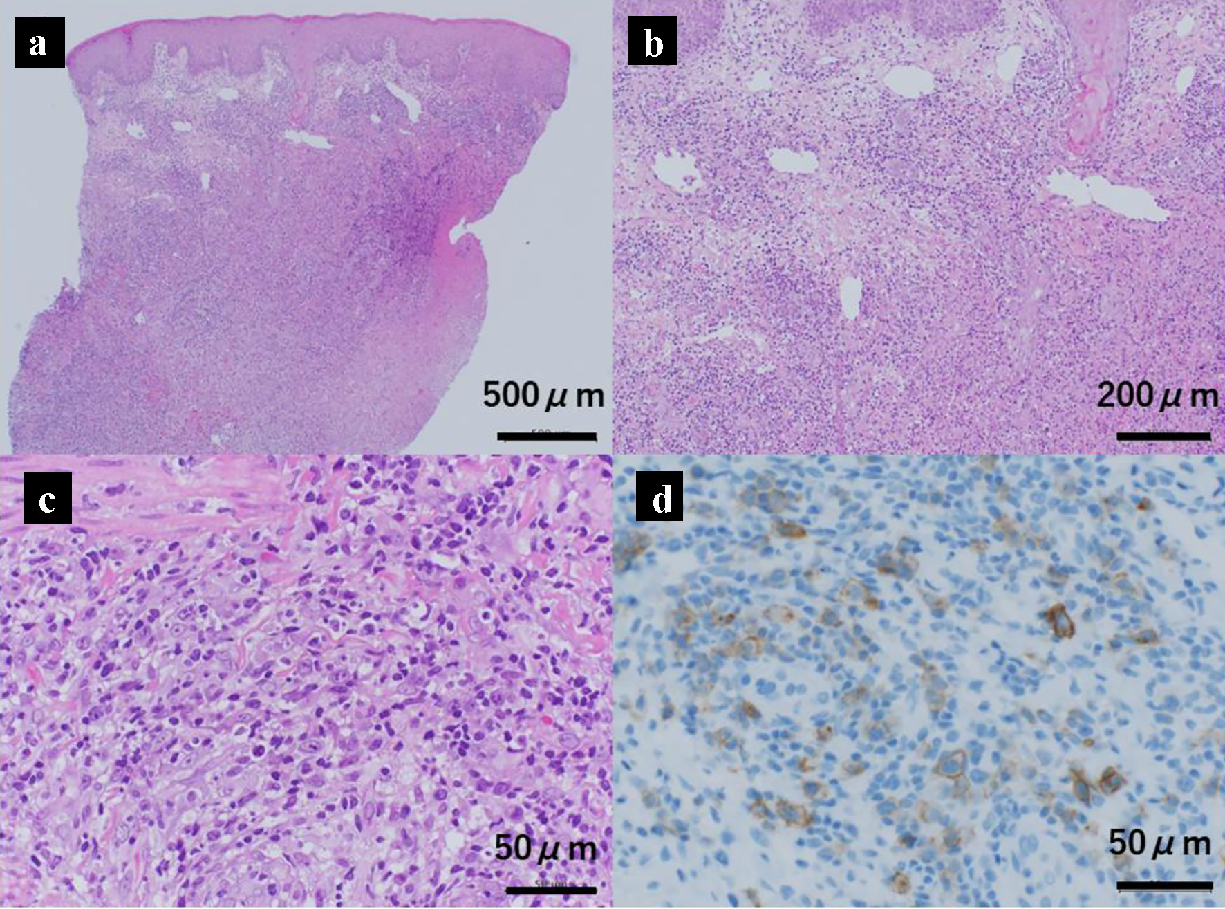
Figures