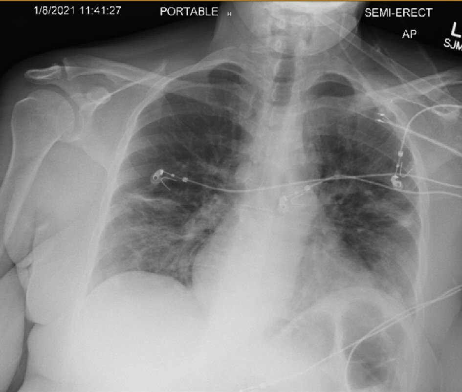
Figure 1. Chest X-ray (single view) showing bilateral ill-defined low-density opacities of mid and lower lung concerning for multifocal viral pneumonia, suggestive of COVID-19 pneumonia. COVID-19: coronavirus disease 2019.
| Journal of Medical Cases, ISSN 1923-4155 print, 1923-4163 online, Open Access |
| Article copyright, the authors; Journal compilation copyright, J Med Cases and Elmer Press Inc |
| Journal website https://www.journalmc.org |
Case Report
Volume 12, Number 8, August 2021, pages 328-331
Ogilvie Syndrome and COVID-19 Infection
Figures