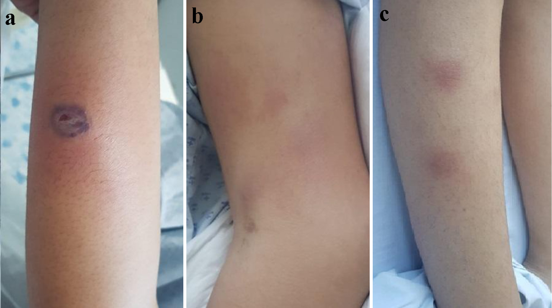
Figure 1. (a) One centimeter purplish-based blister in the middle-third of left forearm. (b) Erythematous lesions in the middle and distal-third of left forearm. (c) Erythematous subcutaneous nodules in the middle-third of right leg.
| Journal of Medical Cases, ISSN 1923-4155 print, 1923-4163 online, Open Access |
| Article copyright, the authors; Journal compilation copyright, J Med Cases and Elmer Press Inc |
| Journal website https://www.journalmc.org |
Case Report
Volume 12, Number 12, December 2021, pages 469-473
Sweet Syndrome in an Adolescent Patient With Differentiation Syndrome Secondary to Promyelocytic Leukemia Treatment With All-Trans Retinoic Acid
Figures
Tables
| BCG: bacille Calmette-Guerin. | |
| Paraneoplastic | Acute lymphoid leukemia |
| Acute myeloid leukemia | |
| Juvenile myelomonocytic leukemia | |
| Myelodysplastic syndromes | |
| Osteosarcoma | |
| Fanconi anemia | |
| Aplastic anemia | |
| Inflammatory diseases | Systemic lupus erythematosus |
| Auto immune hepatitis | |
| Crohn’s disease | |
| Infections | Human immunodeficiency virus |
| Rotavirus | |
| Otitis media | |
| Tonsilitis | |
| Acute respiratory infections | |
| Drug treatment | Colony stimulating factors |
| Trimethoprim sulfamethoxazol | |
| Retinoids | |
| Azathioprine | |
| Contraceptives | |
| Minocycline | |
| Carbamazepine | |
| Tyrosine kinase inhibitors | |
| Vaccines | BCG |
| Measles | |
| Influenza | |
| Pneumococcus | |
| Pregnancy | - |
| ESR: erythrocyte sedimentation rate; PBS: peripheral blood smear; PMN: polymorphonuclear. | |
| Major criteria | Sudden appearance of painful or erythematous plaques, nodules, pustules, or blisters |
| Neutrophilic infiltration of the dermis without leukocytoclastic vasculitis | |
| Minor criteria | Clinical picture preceded by the application of vaccines or a respiratory or gastrointestinal infection, associated or not with: 1) inflammatory diseases such as autoimmune disorders; 2) lymphoproliferative diseases or solid tumors; and 3) pregnancy. |
| Clinical picture preceded by fever and general discomfort | |
| Laboratories results altered at the beginning of the clinical picture: ESR > 20 mm/h, positive C-reactive protein, PBS > 70% PMN, leukocytes > 8,000 cells/µL | |
| Excellent response to treatment with systemic steroids or potassium iodide | |