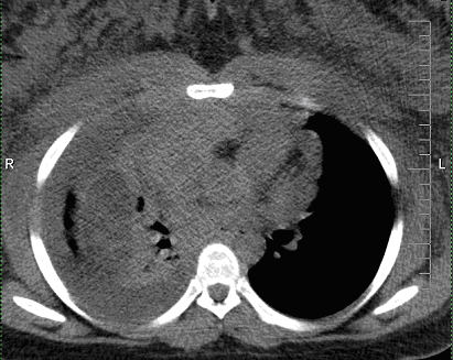
Figure 1. Non-contrasted CT thorax demonstrates large anterior mediastinal mass with associated bulky mediastinal and axillary lymphadenopathies, large right pleural effusion, and anasarca of upper chest wall and breasts.
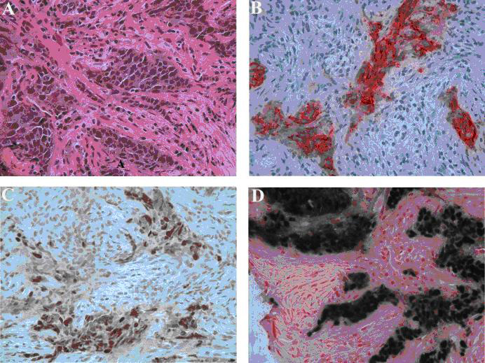
Figure 2. Left axillary lymph node biopsy. (A) Poorly differentiated carcinoma with squamous features (hematoxylin-eosin stain, original magnification, x 400). (B) Tumor positive for cytokeratin (Immunostain for cytokeratin AE1/3, original magnification x 400). (C) Positive nuclear staining for P63 (Immunostain for P63, original magnification x 400) and (D) In-situ hybridization for Epstein-Barr virus shows strong positivity on tumor cells (EBER, original magnification x 400).
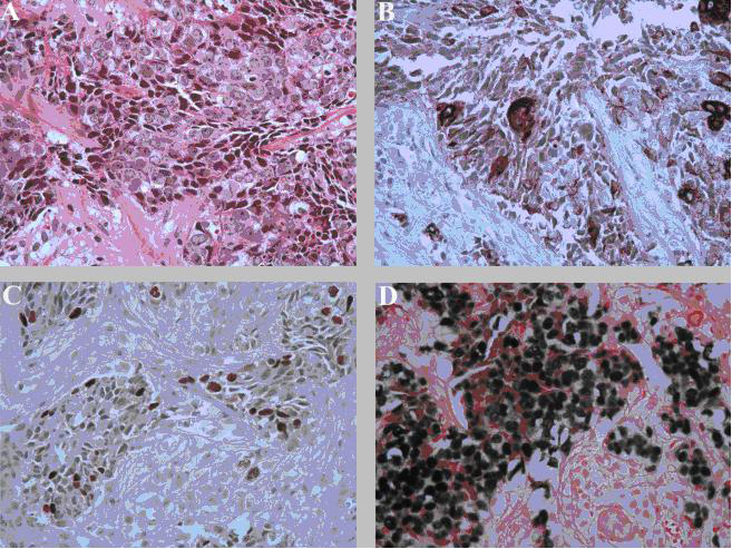
Figure 3. Anterior mediastinal mass biopsy. (A) Poorly differentiated carcinoma with squamous features (hematoxylin-eosin stain, original magnification, x 400). (B) Tumor positive for cytokeratin (Immunostain for pan-cytokeratin, original magnification x 400). (C) Positive nuclear staining for P63 (Immunostain for P63, original magnification x 400) and (D) In-situ hybridization for Epstein-Barr virus shows strong positivity on tumor cells (EBER, original magnification x 400).