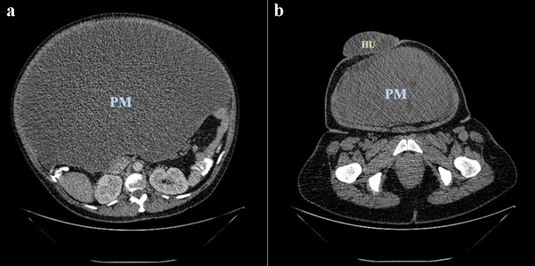
Figure 1. Pelvic mass (PM) (a) and umbilical hernia (HU) (b) seen on abdomen-pelvis computed tomography (CT) scan.
| Journal of Medical Cases, ISSN 1923-4155 print, 1923-4163 online, Open Access |
| Article copyright, the authors; Journal compilation copyright, J Med Cases and Elmer Press Inc |
| Journal website https://www.journalmc.org |
Case Report
Volume 12, Number 10, October 2021, pages 386-390
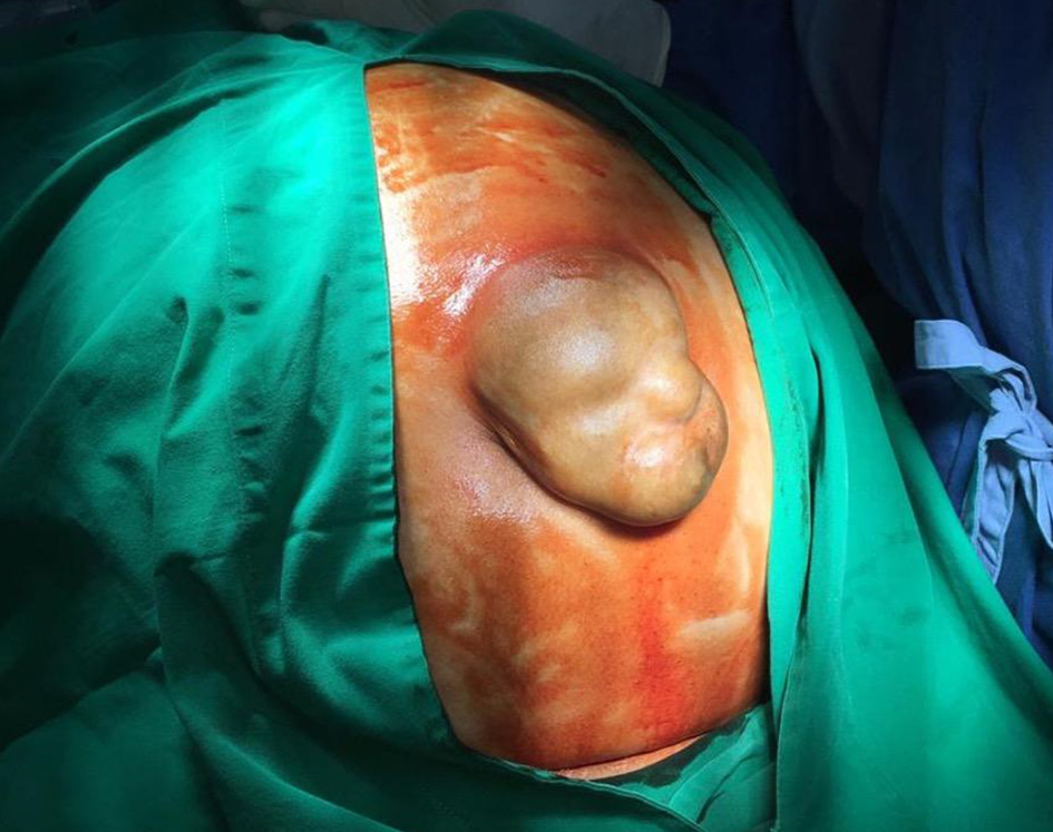
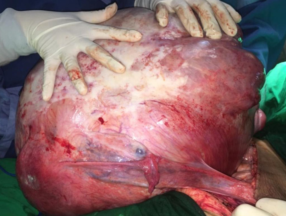
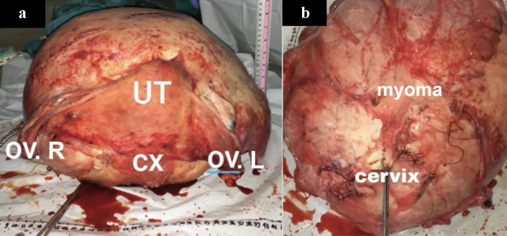
Giant Uterine Leiomyoma With Surgical Difficulty
Figures