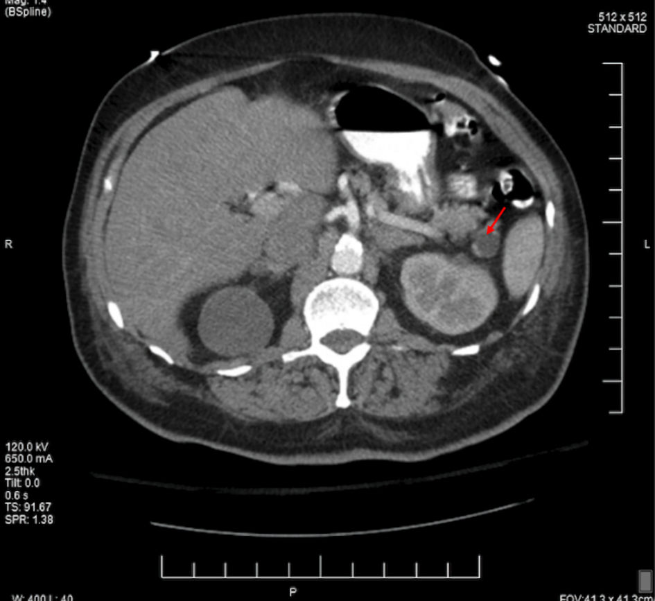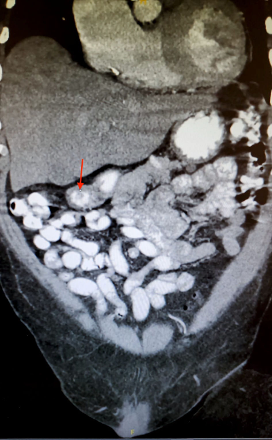
Figure 1. The CT scan in coronal view of the abdomen showcasing 1.3-cm duodenal filling defect (red arrow). CT: computed tomography.
| Journal of Medical Cases, ISSN 1923-4155 print, 1923-4163 online, Open Access |
| Article copyright, the authors; Journal compilation copyright, J Med Cases and Elmer Press Inc |
| Journal website https://www.journalmc.org |
Case Report
Volume 12, Number 10, October 2021, pages 419-423
A Rare Case of Three Distinct Gastrointestinal Neoplasms Occurring Simultaneously in an Elderly Patient
Figures