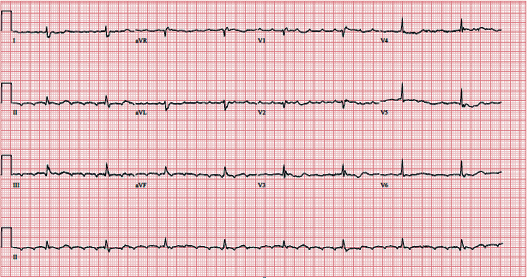
Figure 1. Atrial flutter with 4:1 block.
| Journal of Medical Cases, ISSN 1923-4155 print, 1923-4163 online, Open Access |
| Article copyright, the authors; Journal compilation copyright, J Med Cases and Elmer Press Inc |
| Journal website https://www.journalmc.org |
Case Report
Volume 12, Number 11, November 2021, pages 433-437
Primary Cardiac Lymphoma: A Case Report
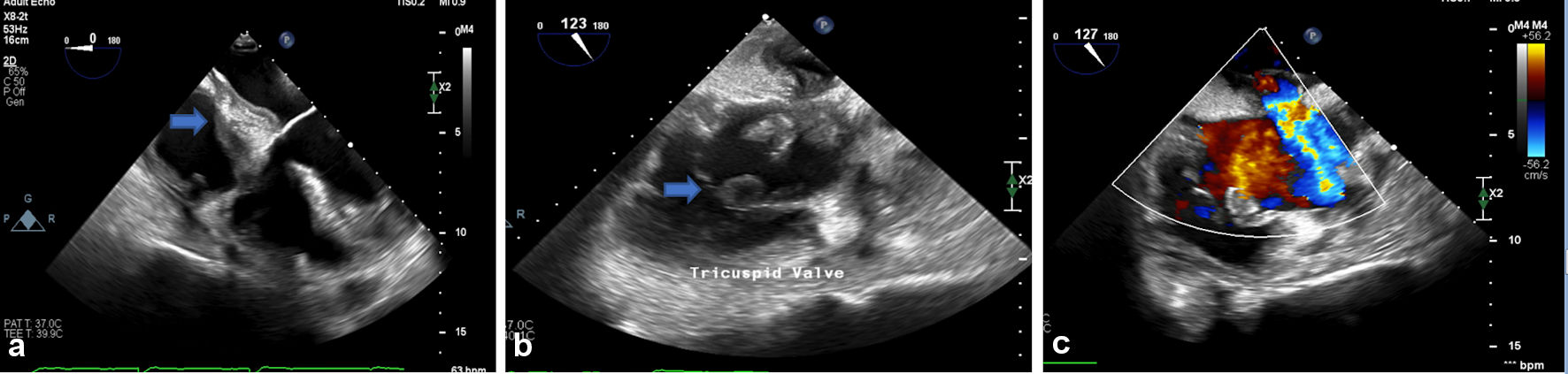
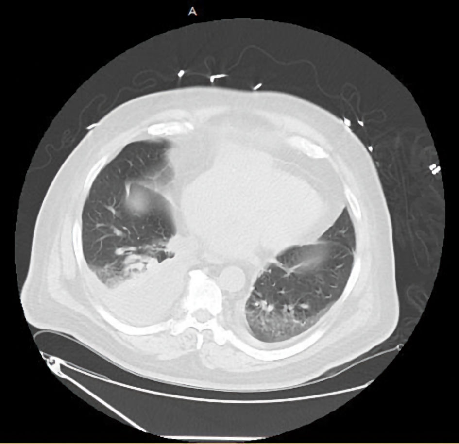
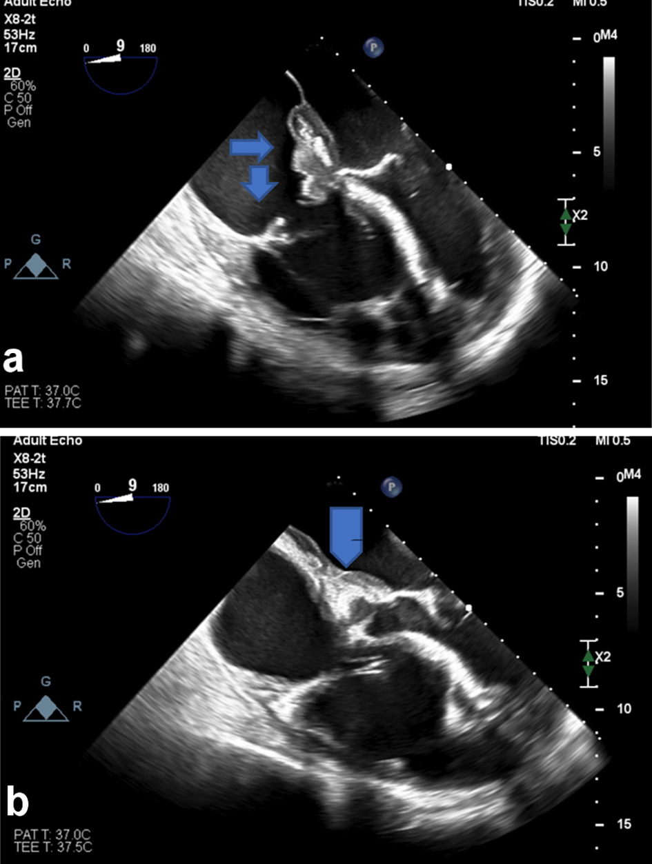
Figures