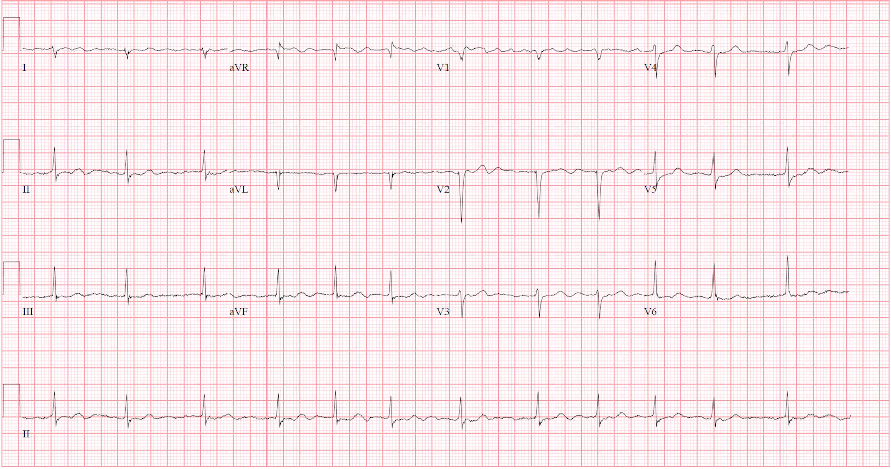
Figure 1. Presenting electrocardiogram showing atrial fibrillation with rate to 75 beats per minute, without ST-T changes.
| Journal of Medical Cases, ISSN 1923-4155 print, 1923-4163 online, Open Access |
| Article copyright, the authors; Journal compilation copyright, J Med Cases and Elmer Press Inc |
| Journal website https://www.journalmc.org |
Case Report
Volume 12, Number 11, November 2021, pages 460-463
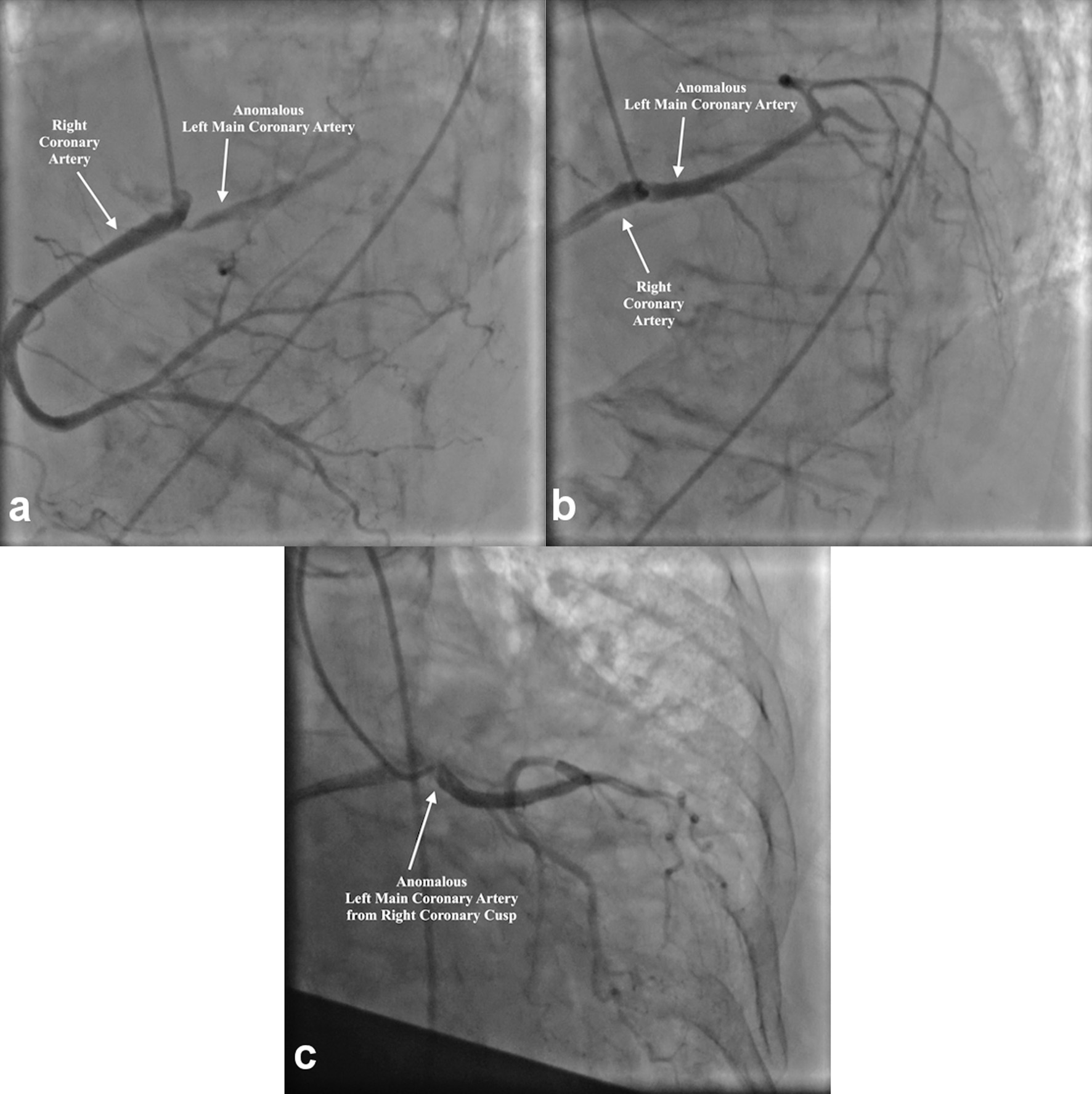
Anomalous Coronary Arteries: A Case of Rare and Incidental Findings
Figures
Table
| Laboratory | Results | References |
|---|---|---|
| Hemoglobin (g/dL) | 11.5 | 12.0 - 16 |
| White blood cell count (× 109/L) | 10.2 | 4.5 - 11.0 |
| Sodium (mmol/L) | 134 | 135 - 146 |
| Potassium (mmol/L) | 5.5 | 3.5 - 5.0 |
| Glucose (mg/dL) | 134 | 70 - 110 |
| Blood urea nitrogen (mg/dL) | 37 | 7.0 - 18.0 |
| Creatinine (mg/dL) | 1.15 | 0.44 - 1.0 |
| B-type natriuretic peptide (pg/mL) | 719 | 0 - 100 |
| Aspartate aminotransferase (U/L) | 62 | 10 - 42 |
| Alanine aminotransferase (U/L) | 30 | 10 - 60 |
| Total bilirubin (mg/dL) | 1.8 | 0.2 - 1.2 |