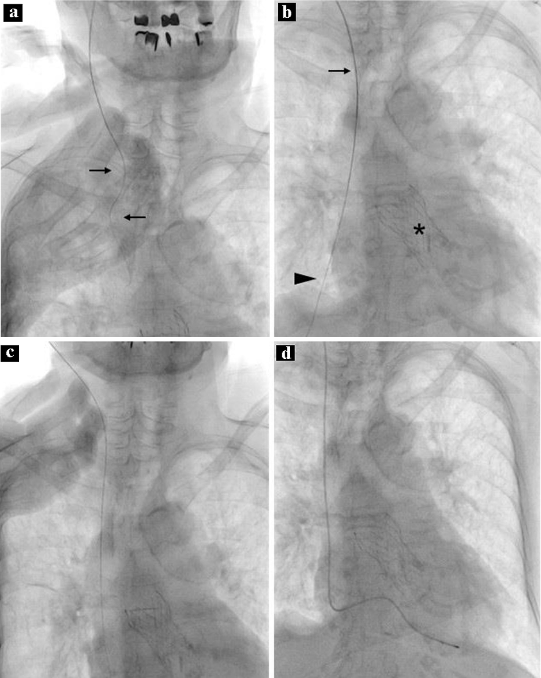
Figure 1. Procedure during temporary pacing. Note that the wire was not straight (a, arrows). A sheath was proceeded along the guidewire; the top of the sheath is in the upper part of the superior vena cava (b, arrow). Note that the distal part of the guidewire was not in the shadow of the heart (b, arrowhead). The asterisk shows an artificial aortic valve. After removal of the sheath and guidewire, the guidewire is correctly inserted into the superior vena cava, right atrium, and inferior vena cava (c). A temporary pacing lead is placed at the apex of the right ventricle (d).