Journal of Medical Cases, ISSN 1923-4155 print, 1923-4163 online, Open Access
Article copyright, the authors; Journal compilation copyright, J Med Cases and Elmer Press Inc
Journal website https://www.journalmc.org

Case Report

Volume 12, Number 11, November 2021, pages 442-445


Cancer of Unknown Primary With Presumptive Pancreatic Cancer Treated With FOLFIRINOX

Figures

Figure 1.
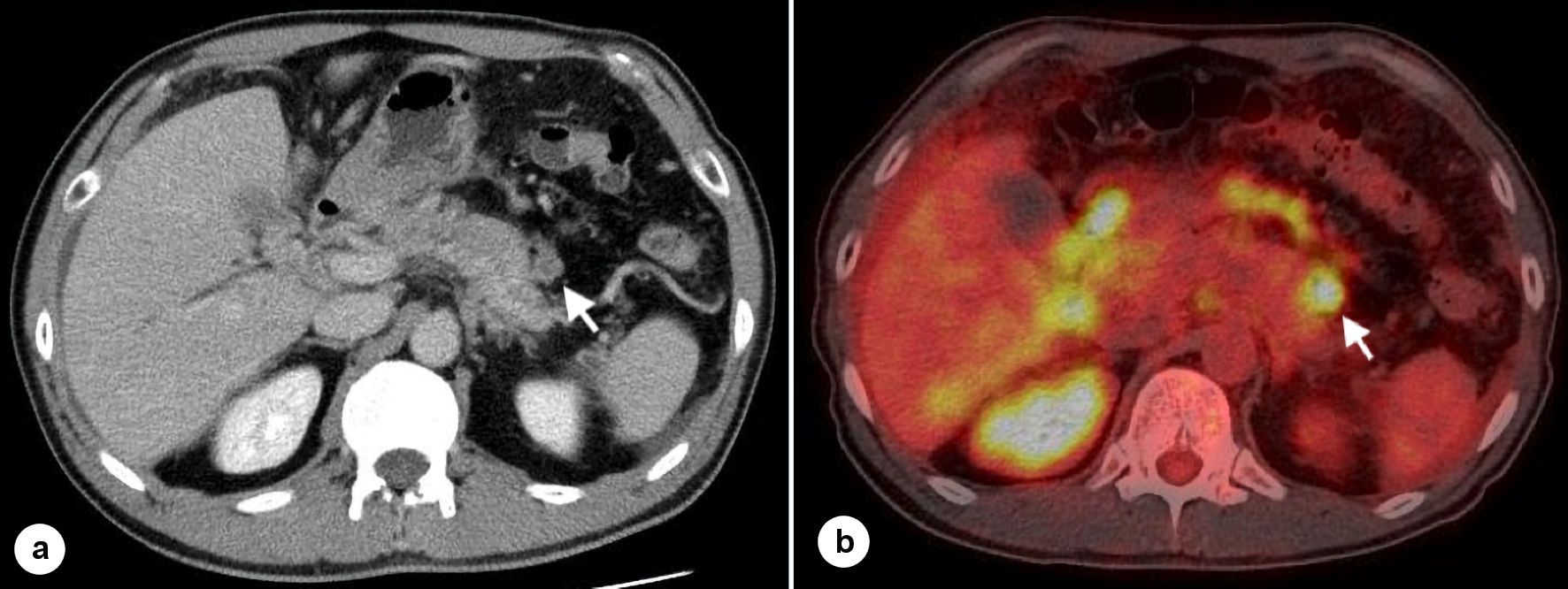
Figure 1. CT scan and PET-CT scan. (a) Intravenous contrast-enhanced CT scans show the soft tissue shadow near the tail of the pancreas (arrow). (b) PET-CT scans show the soft tissue shadow near the tail of the pancreas (arrow). CT: computed tomography; PET: positron emission tomography.
Figure 2.
Figure 2. EUS-FNA of the soft tissue shadow near the tail of the pancreas. EUS showing the soft tissue shadow near the tail of the pancreas (arrow head) is punctured by the needle (arrow). EUS-FNA: endoscopic ultrasound/fine needle aspiration.
Figure 3.
Figure 3. Neoplastic cells, hematoxylin-eosin stain, immunohistochemical stains × 100. (a) Hematoxylin-eosin staining of neoplastic cells. (b) Immunohistochemical stain of CK7 showing positivity in neoplastic cells. (c) Immunohistochemical stain of CK20 showing positivity in neoplastic cells (scattered). (d) Immunohistochemical stain of CA19-9 showing positivity in neoplastic cells. CK: cytokeratin; CA: carbohydrate antigen.