Journal of Medical Cases, ISSN 1923-4155 print, 1923-4163 online, Open Access
Article copyright, the authors; Journal compilation copyright, J Med Cases and Elmer Press Inc
Journal website https://www.journalmc.org

Case Report

Volume 12, Number 12, December 2021, pages 516-519


Idiopathic Sclerosing Mesenteritis: An Extremely Rare Cause of Mesenteric Mass

Figures

Figure 1.
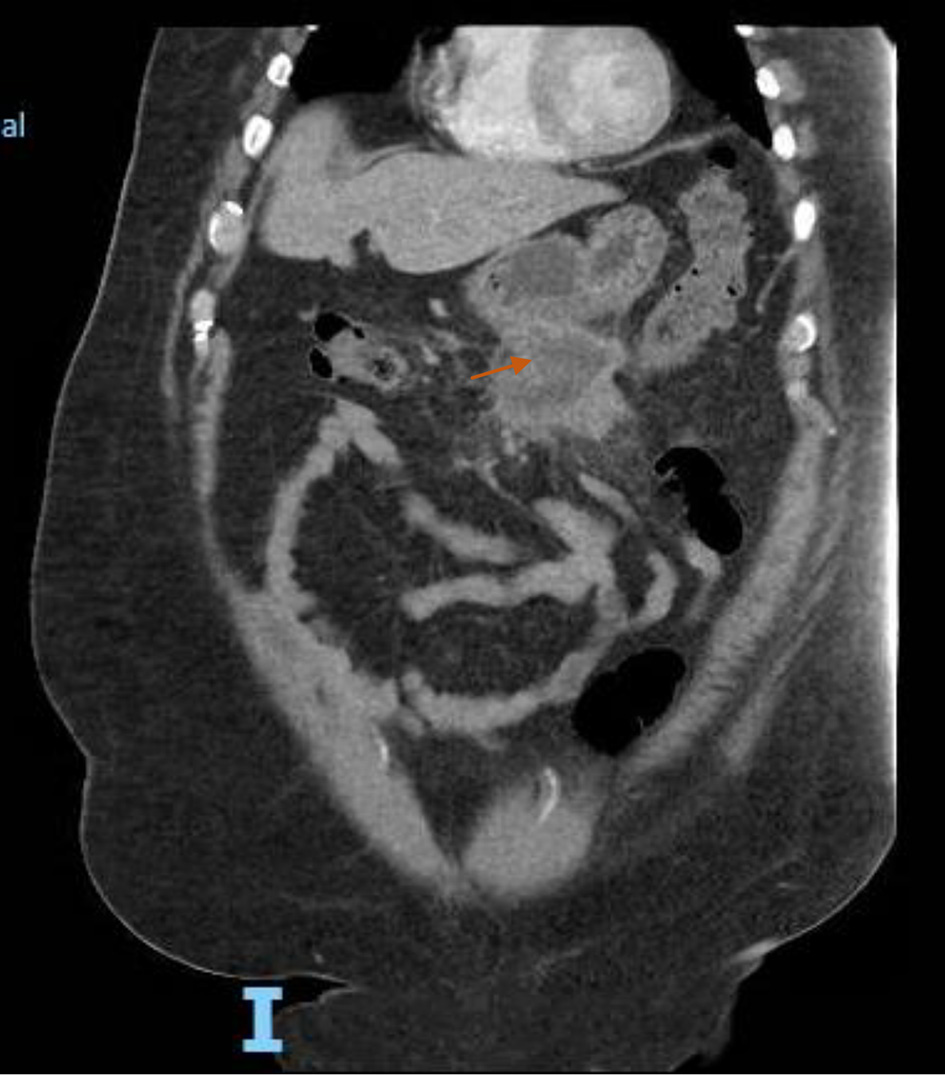
Figure 1. A coronal section of abdomen computed tomography with intravenous contrast demonstrating a rim enhancing hypodense amorphous lesion in mesentery. This lesion is contiguous with the inferior aspect of the greater curvature of the stomach as well as a loop of bowel. Also surrounding the lesion, an extensive fat stranding was noted (orange arrow).
Figure 2.
Figure 2. A Linear echo endoscopic image showing irregular hypoechoic mass of size 3.7 cm (orange arrow) outside the gastric echo layer in distal gastric body.
Figure 3.
Figure 3. (a) Fat necrosis, area of fibrosis and infiltration of chronic inflammatory cells (H&E, magnification, × 20). (b) Infiltration of lipid laden macrophages (H&E, magnification, × 40). H&E: hematoxylin and eosin stain.