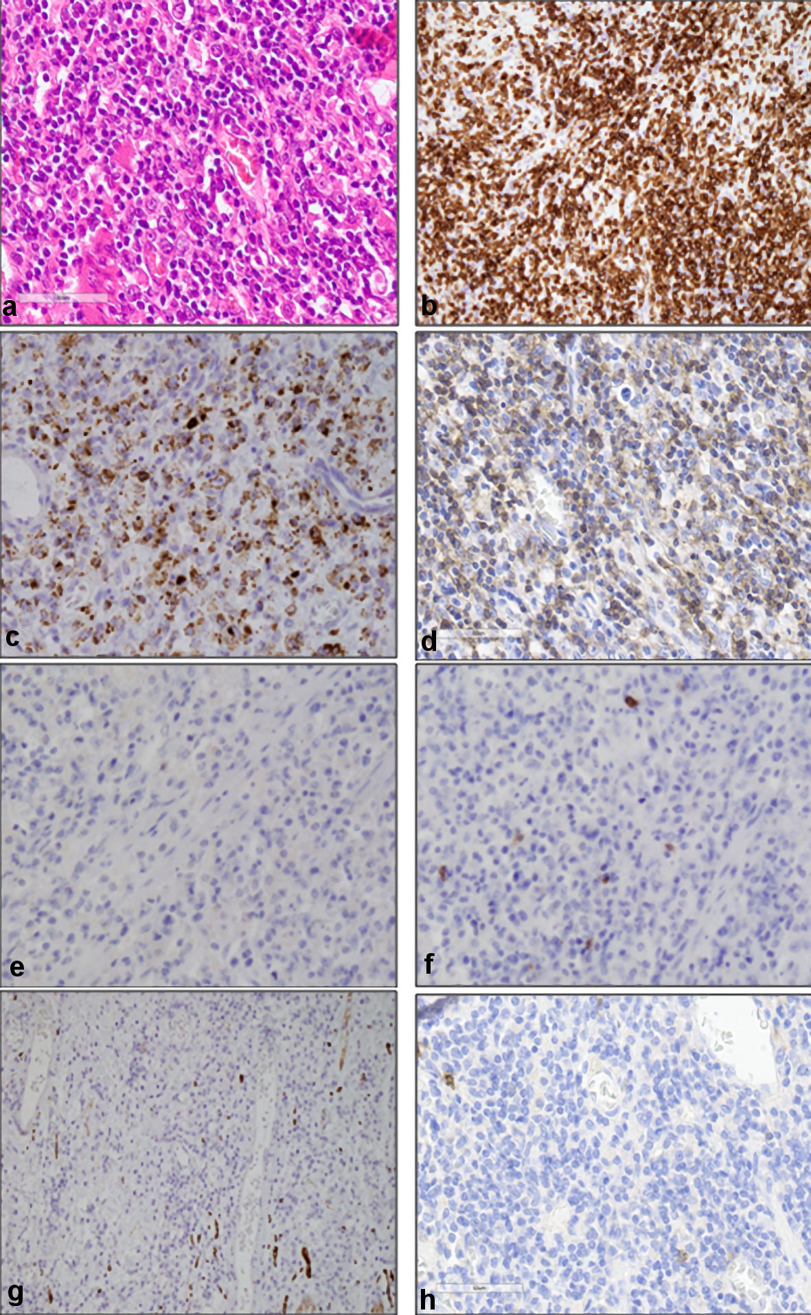
Figure 1. Histopathological findings of resected colon. (a) Dense pleomorphic medium-sized abnormal lymphocytes, as well as infiltrating inflammatory cells (hematoxylin and eosin staining, original magnification, × 400). Immunohistochemical analysis showed that medium-sized abnormal lymphocytes were positive for CD3 (b), granzyme B (c), and TCR-βF1 (d), and negative for CD4 (e), CD8 (f), CD56 (g), and CD103 (h) (original magnification, × 400). CD: cluster of differentiation; TCR: T cell receptor.
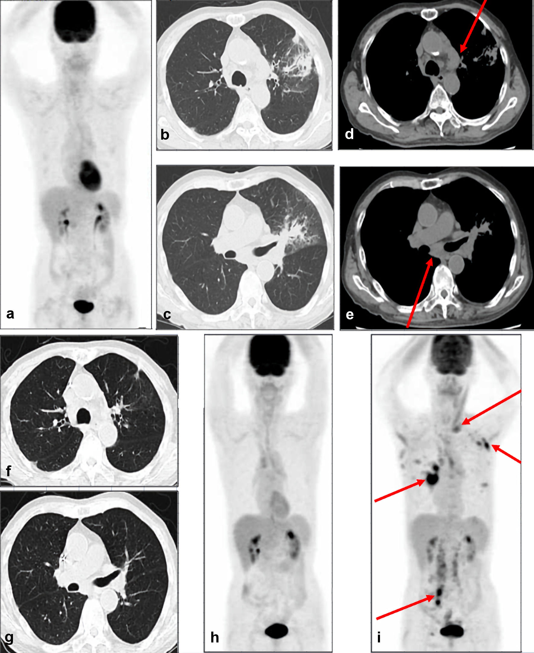
Figure 2. CT and PET-CT images. (a) PET-CT images show no abnormal accumulation of fluorodeoxyglucose in the whole body, suggesting CMR. CT images at relapse show infiltration and ground-glass opacity in the left pulmonary area (b, c), in addition to the enlargement of mediastinal and left subclavian lymph nodes (d, e). Arrows indicate enlarged lymph nodes. CT images after three courses of GDP chemotherapy show the disappearance of pulmonary shadows and lymphadenopathy (f, g). (h) PET-CT images after three courses of GDP chemotherapy show no abnormal accumulation of fluorodeoxyglucose in the whole body, suggesting CMR. (i) PET-CT images at second relapse show the accumulation of fluorodeoxyglucose in multiple lymph nodes in left supraclavicular fossa, left axilla, bilateral hilum, mesentery, para-aorta, right iliac area, and right inguinal area. Arrows indicate the abnormal accumulation in lymph nodes. PET-CT: positron emission tomography-computed tomography; GDP: gemcitabine, dexamethasone, and cisplatin; CMR: complete metabolic remission.
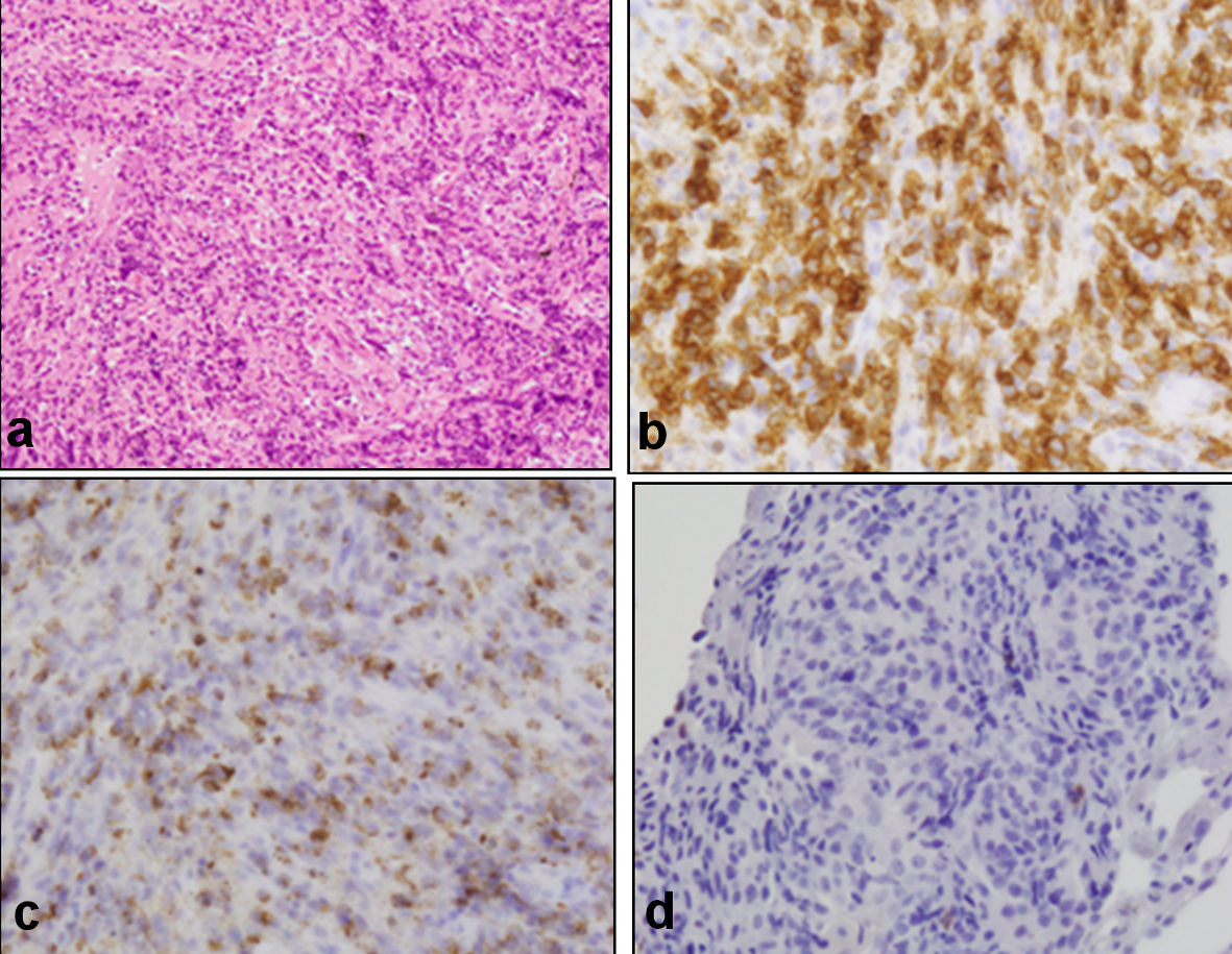
Figure 3. Histopathological findings of the biopsied samples from lung. (a) Medium-sized abnormal lymphocytes densely occupying alveolar septum, as well as fibrosis and localized thickening of the alveolar septum (hematoxylin and eosin staining, original magnification, ×200). Immunohistochemical analysis showed that medium-sized abnormal lymphocytes were positive for CD3 (b) and granzyme B (c) and negative for CD8 (d), consistent with relapsed ITL, NOS (original magnification, ×400). CD: cluster of differentiation; NOS: not otherwise specified.