Journal of Medical Cases, ISSN 1923-4155 print, 1923-4163 online, Open Access
Article copyright, the authors; Journal compilation copyright, J Med Cases and Elmer Press Inc
Journal website https://www.journalmc.org

Case Report

Volume 13, Number 4, April 2022, pages 183-187


Endoscopic Ultrasound-Guided Rendezvous for Biliary Obstruction in Patient With Prior Whipple Surgery

Figures

Figure 1.
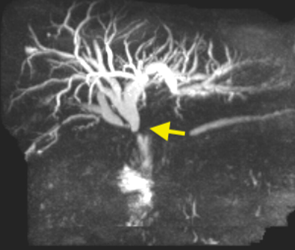
Figure 1. Magnetic resonance cholangiopancreatography showing abrupt cut off at the level of the hepaticojejunal anastomosis (arrow) with diffuse biliary dilation.
Figure 2.
Figure 2. Suture material seen in jejunum suggestive of the biliary (afferent) limb.
Figure 3.
Figure 3. Endoscopic ultrasound showing left intra-hepatic biliary radical (arrow).
Figure 4.
Figure 4. Contrast injection via echoendoscope showing diffuse dilation of biliary tree with abrupt cut off at the hepaticojejunal anastomosis (arrow).
Figure 5.
Figure 5. Fluoroscopy showing wire passage across the hepaticojejunal anastomosis into the jejunum (long yellow arrow), with short red arrows highlighting the view of hepaticojejunal anastomosis from the surgical clips.
Figure 6.
Figure 6. (a) Fluoroscopic view showing cannulation of the hepaticojejunal anastomosis using the modified therapeutic gastroscope (arrow). (b) Endoscopic view post placement of uncovered metal stent across the hepaticojejunal anastomosis (right).