Journal of Medical Cases, ISSN 1923-4155 print, 1923-4163 online, Open Access
Article copyright, the authors; Journal compilation copyright, J Med Cases and Elmer Press Inc
Journal website https://www.journalmc.org

Case Report

Volume 13, Number 2, February 2022, pages 66-70


Rare Presentation of Mycobacterium tuberculosis Mimicking Prostate Cancer

Figures

Figure 1.
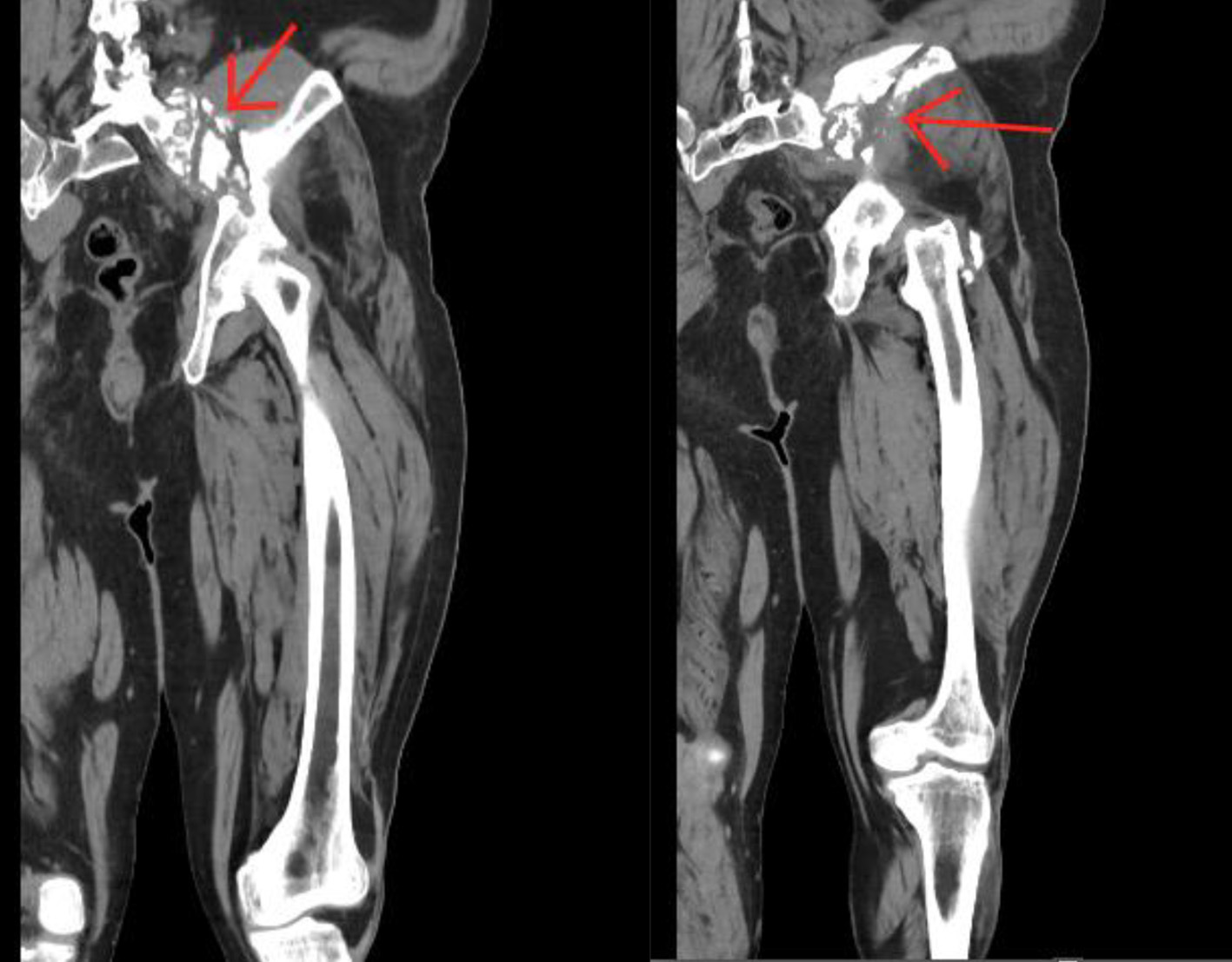
Figure 1. Coronal computer tomography scan of the lower extremity identifying the lesion on the left hip (arrows).
Figure 2.
Figure 2. Axial computer tomography scan of the lower extremity identifying the lesion on the left hip (arrows).
Figure 3.
Figure 3. (a) H&E slide (low magnification, × 4) shows necrotic and viable tissue with non-necrotizing granuloma; (b) H&E slide (low magnification, × 10) shows granuloma and necrotic tissue; (c) H&E slide (low magnification, × 10) of well-formed granulomas showing the epithelioid histiocytes and multinucleated giant cells; (d) H&E slide (high magnification, × 40) of a granuloma showing epithelioid histiocytes and multinucleated giant cells. H&E: hematoxylin and eosin.