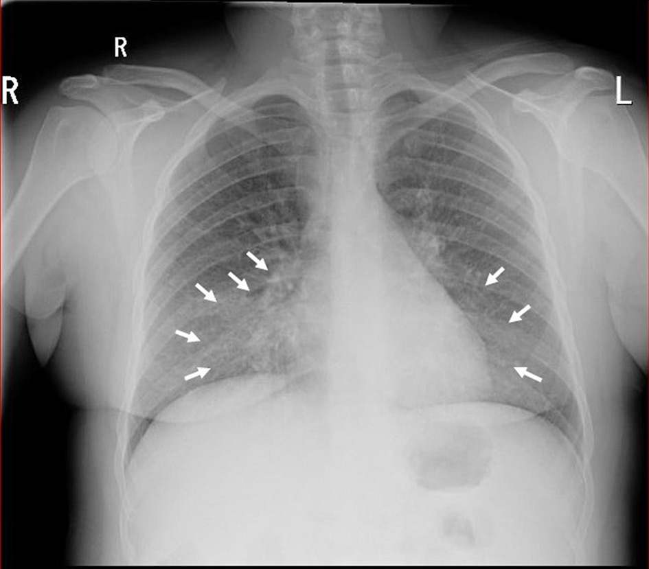
Figure 1. Chest X-ray demonstrating right and left mid-zone opacification consistent with bilateral pneumonia (white arrows).
| Journal of Medical Cases, ISSN 1923-4155 print, 1923-4163 online, Open Access |
| Article copyright, the authors; Journal compilation copyright, J Med Cases and Elmer Press Inc |
| Journal website https://www.journalmc.org |
Case Report
Volume 13, Number 3, March 2022, pages 119-124
A Rare Case of Severe Hemolytic Anemia and Pulmonary Embolism Secondary to Mycoplasma pneumoniae Infection
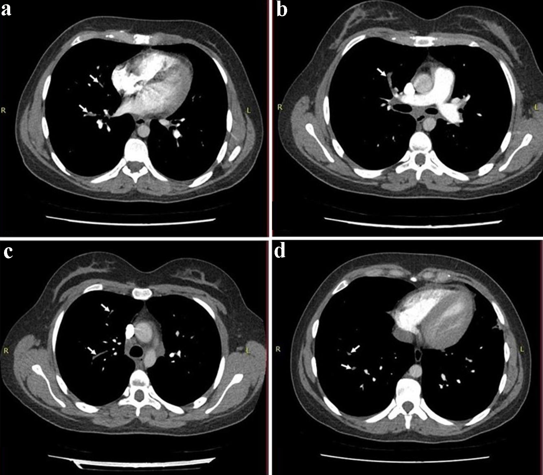
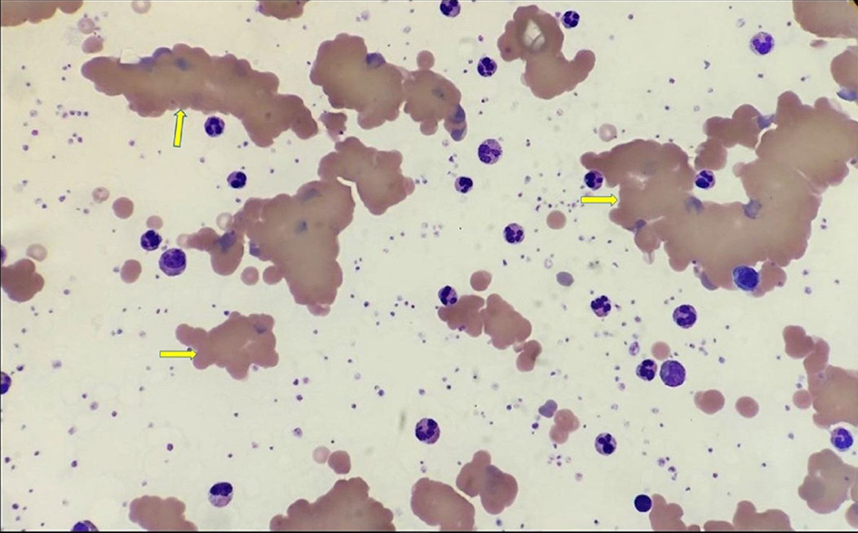
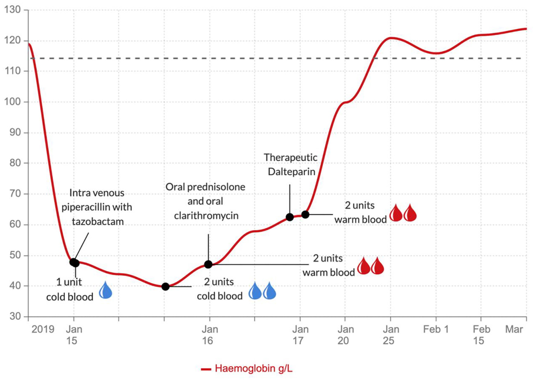
Figures