Journal of Medical Cases, ISSN 1923-4155 print, 1923-4163 online, Open Access
Article copyright, the authors; Journal compilation copyright, J Med Cases and Elmer Press Inc
Journal website https://www.journalmc.org

Case Report

Volume 13, Number 3, March 2022, pages 109-114


A Case of Angioimmunoblastic T-cell Lymphoma Hidden in Plain Sight: A Delay in Diagnosis and Management

Figures

Figure 1.
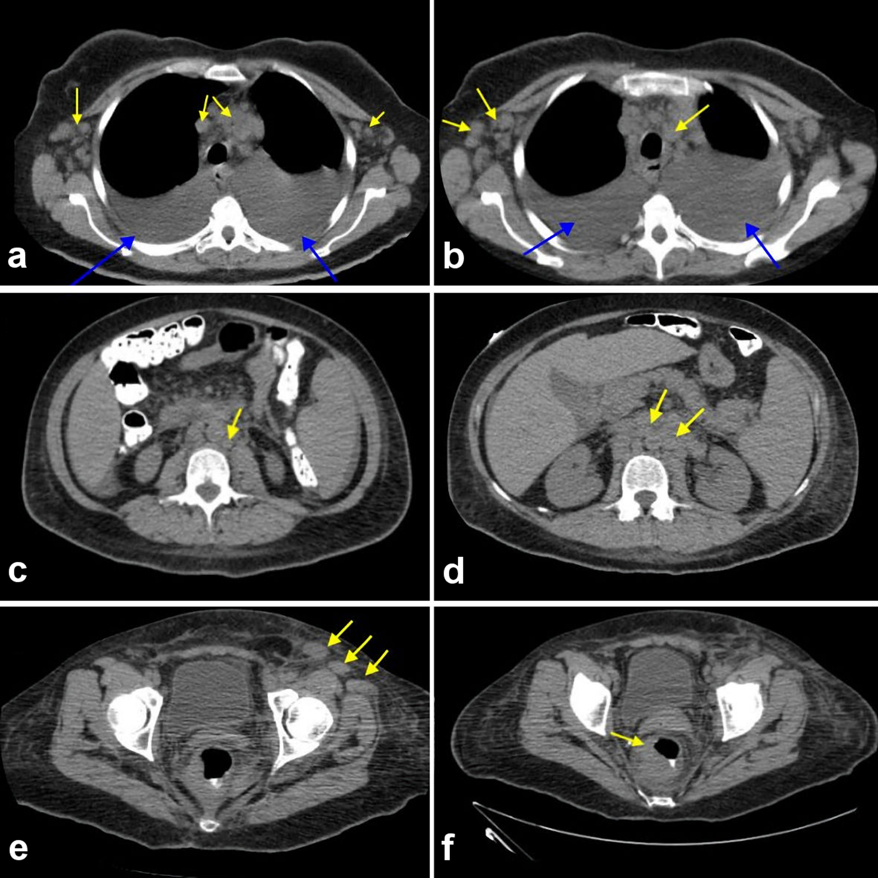
Figure 1. (a-f) Computed tomography scan of chest, abdomen, and pelvis showing an extensive lymphadenopathy in the bilateral axillary region and in the mediastinum (yellow arrows) with bilateral pleural effusion noted (blue arrows). (a) Extensive lymphadenopathy in bilateral axillary region and in the mediastinum (yellow arrows). (b) Extensive lymphadenopathy involving the bilateral thoracic region and mediastinum (yellow arrows). (c) Extensive paraaortic and pre-vertebral lymphadenopathy (yellow arrows). (d) Extensive paraaortic wall lymphadenopathy (yellow arrows). (e) Extensive pelvic wall lymphadenopathy. (f) Discrete mass surrounding the rectum suspicious for malignancy.
Figure 2.
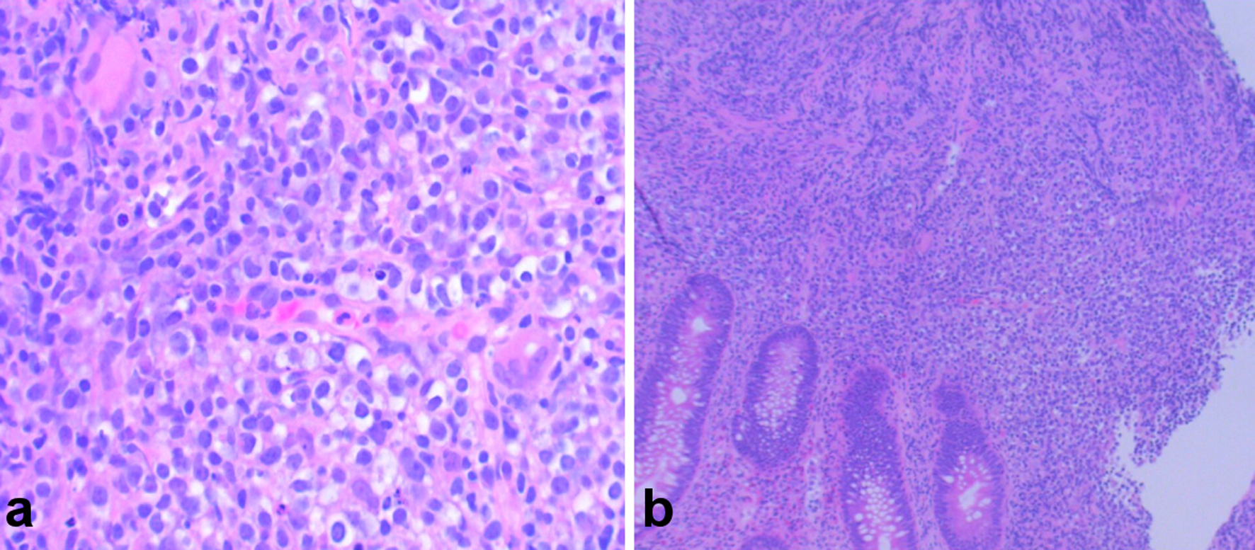
Figure 2. (a, b) Hematoxylin and eosin (H&E) stain showing colonic mucosa with atypical proliferation of atypical lymphocytes, consistent with angioimmunoblastic T-cell lymphoma (AITL).
Figure 3.
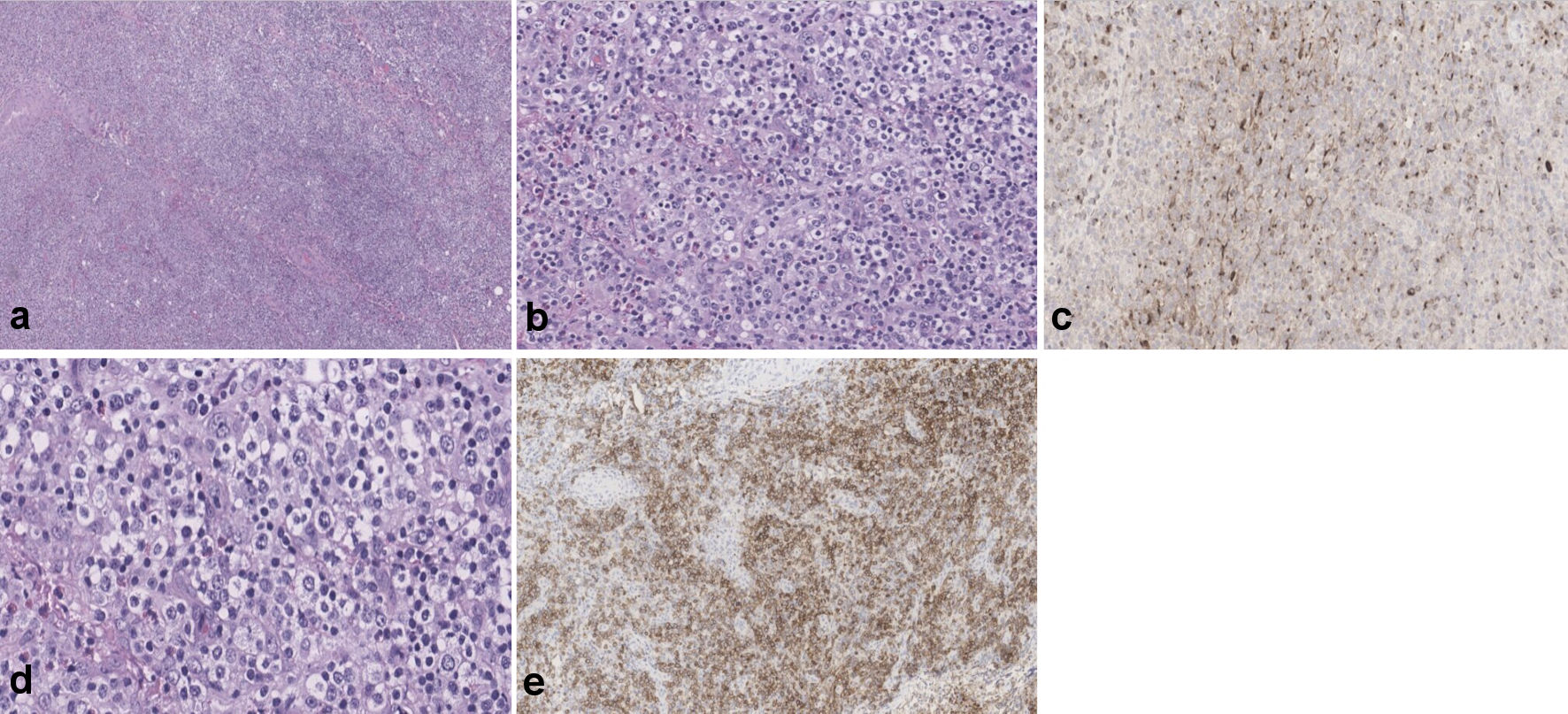
Figure 3. (a-c) Showing portions of lymphoid tissue with effacement of normal architecture by largely diffuse, polymorphic infiltrates of small- to medium-sized lymphocytes (frequently grouped showing clear cytoplasm), scattered large, transformed cells, many eosinophils and plasma cells. Background prominent vasculature, composed of arborizing high endothelial venules is evident. Large, atypical lymphocytes are positive for ICOS and CXCL-13 (d, e). ICOS: inducible T-cell co-stimulator; CXCL-13: chemokine (CXC motif) ligand 13.

Table

Table 1. Laboratory Studies Done During Admission
 
ResultsReference range
HIV: human immunodeficiency virus; HTLV: human T-lymphotropic virus; COVID-19: coronavirus disease 2019; SARS-CoV-2: severe acute respiratory syndrome coronavirus 2.
Blood, plasma, serum
  Hemoglobin (g/dL)10.312.0 - 16.0
  White blood cells (× 103/µL)3.24.5 - 11.0
  Platelet count (× 103/µL)222140 - 450
  Sodium (mmol/L)141136 - 145
  Potassium (mmol/L)4.23.5 - 5.0
  Glucose (mg/dL)11670 - 99
  Albumin (g/dL)2.83.5 - 5.0
  Total protein (g/dL)4.86 - 8
  Blood urea nitrogen (BUN) (mg/dL)95 - 25
  Creatinine (mg/dL)0.750.44 - 1.0
  Lactate dehydrogenase (U/L)23291 - 200
  Haptoglobin (mg/dL)13930 - 225
  Erythrocyte sedimentation rate (mm/h)630 - 30
  Vitamin B12 (pg/mL)79180 - 914
  HIV Ag/Ab 4th generationNot detectedNot detected
  HTLV 1/2 antibody screenNon-reactiveNon-reactive
  SARS-COV-2 (COVID-19)NegativeNegative
  Antinuclear antibody (ANA) testPositiveNegative
  ANA titer1:40< 1:40
  Hepatitis acute panelNegativeNegative
Pleural fluid analysis
  AppearanceClearN/A
  ColorYellowN/A
  White blood cells (/µL)8250 - 200
  Red blood cells (/µL)45 - 1500
  Segmented neutrophils (%)40 - 25
  Lymphocytes (%)870
  Eosinophils (%)10
  Mono/macrophage (%)80
  Total protein (g/dL)3.4< 40
  Glucose (mg/dL)11430 - 50
  Lactate dehydrogenase (U/L)22991 - 200