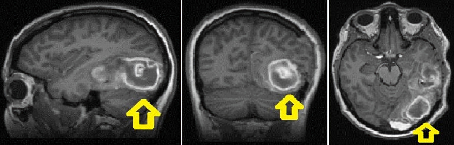
Figure 1. Brain MRI showed left temporal occipital intraparenchymal hematoma (as shown by arrows) measuring 3.4 × 2.4 × 3.2 cm with surrounding edema, and rightward midline shift with subfalcine and uncal herniation. MRI: magnetic resonance imaging.
| Journal of Medical Cases, ISSN 1923-4155 print, 1923-4163 online, Open Access |
| Article copyright, the authors; Journal compilation copyright, J Med Cases and Elmer Press Inc |
| Journal website https://www.journalmc.org |
Case Report
Volume 13, Number 4, April 2022, pages 151-154
A Diagnosis of Choriocarcinoma in a Parturient Presenting With Intracranial Hemorrhage
Figures