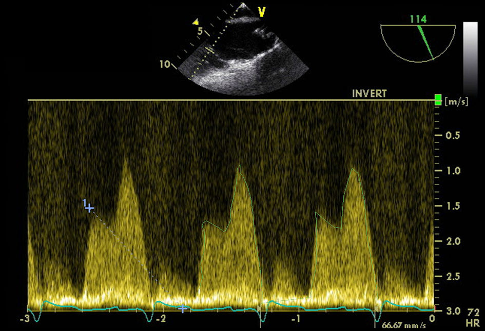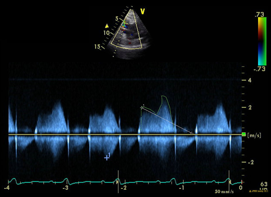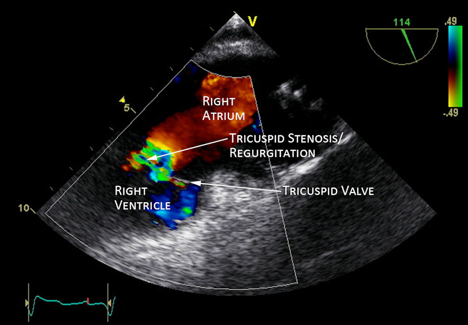
Figure 1. Transthoracic echocardiogram showed tricuspid stenosis and regurgitation (white arrow).
| Journal of Medical Cases, ISSN 1923-4155 print, 1923-4163 online, Open Access |
| Article copyright, the authors; Journal compilation copyright, J Med Cases and Elmer Press Inc |
| Journal website https://www.journalmc.org |
Case Report
Volume 13, Number 8, August 2022, pages 365-368
Severe Tricuspid Stenosis Secondary to Permanent Pacemaker Lead
Figures