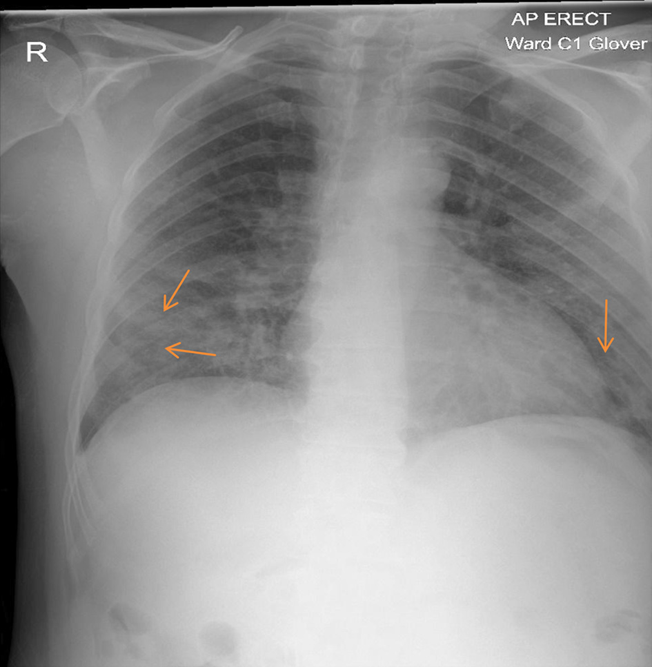
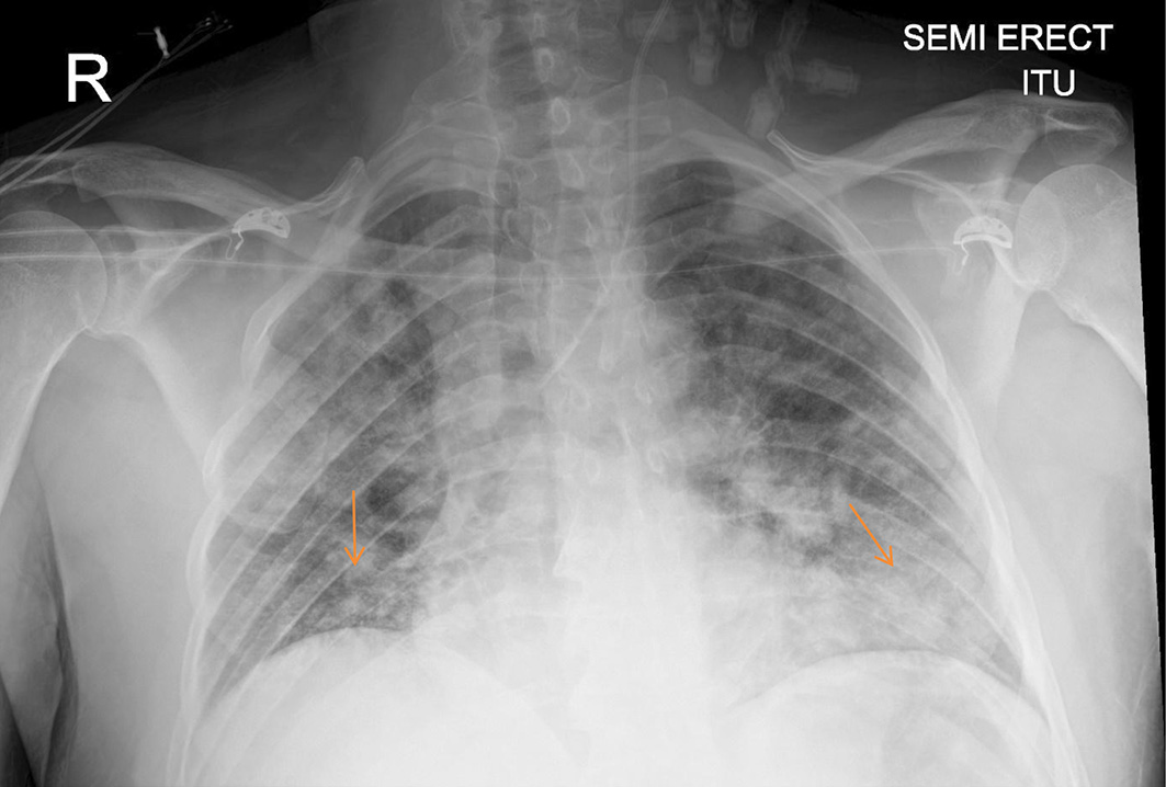
Figure 1. Chest X-ray: increased air space shadowing (arrows) in the mid and lower zones with a peripheral pattern suggestive of COVID-19 pneumonia. COVID-19: coronavirus disease 2019.
| Journal of Medical Cases, ISSN 1923-4155 print, 1923-4163 online, Open Access |
| Article copyright, the authors; Journal compilation copyright, J Med Cases and Elmer Press Inc |
| Journal website https://www.journalmc.org |
Case Report
Volume 13, Number 5, May 2022, pages 207-211
A Case of Delayed COVID-19-Related Macrophage Activation Syndrome
Figures
Table
| Criteria | First admission | Second admission |
|---|---|---|
| LDH: lactate dehydrogenase; AST: aspartate aminotransferase; ALT: alanine aminotransferase; CRP: C-reactive protein. | ||
| Fever (temperature more than 38 °C) | 39.5 °C | 38.2 °C |
| Ferritin concentration (40 - 405 µg/L) | 1,620 | > 99,999 |
| Neutrophil count (2.1 - 7.4 × 10⁹/L) | 12.55 | 11.15 |
| Lymphocytes count (1.0 - 3.6 × 10⁹/L) | 0.59 | 0.45 |
| Platelets count (150 - 400 × 109/L) | 240 | 65 |
| Quantitative D-dimer (0 - 250 ng/mL) | 446 | 375 |
| LDH level (135 - 225 U/L) | 334 | 543 |
| AST level (0 - 40 U/L) | Not done | 23,118 |
| ALT level (0 - 41 U/L) | 101 | 13,685 |
| CRP level (0 - 5 mg/L) | 244 | 55 |
| Triglycerides level (less than 1.7 mmol/L) | 1.3 | 1.9 |