Journal of Medical Cases, ISSN 1923-4155 print, 1923-4163 online, Open Access
Article copyright, the authors; Journal compilation copyright, J Med Cases and Elmer Press Inc
Journal website https://www.journalmc.org

Case Report

Volume 13, Number 7, July 2022, pages 313-317


Two Patients With Difficulty in Swallowing due to Dysphagia Lusoria

Figures

Figure 1.
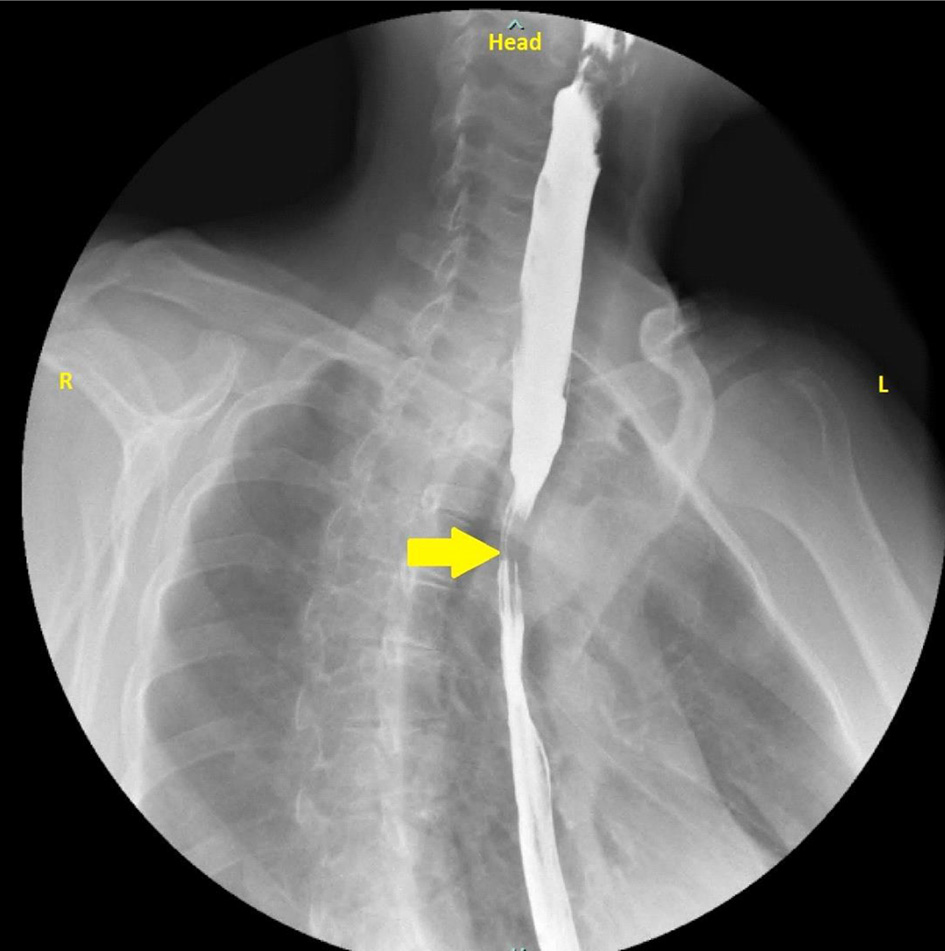
Figure 1. Barium swallow with esophageal narrowing (yellow arrow).
Figure 2.
Figure 2. CT angiography of the chest showing arrow (blue) pointing at the aberrant right subclavian artery compressing the esophagus and the trachea. CT: computed tomography.
Figure 3.
Figure 3. EGD with a normal esophagus and no pulsatile indentation. EGD: esophagogastroduodenoscopy.
Figure 4.
Figure 4. CT angiography showing an ARSA causing external compression of the esophagus with no tracheal compression (arrows). CT: computed tomography; ARSA: aberrant right subclavian artery.