Journal of Medical Cases, ISSN 1923-4155 print, 1923-4163 online, Open Access
Article copyright, the authors; Journal compilation copyright, J Med Cases and Elmer Press Inc
Journal website https://www.journalmc.org

Case Report

Volume 13, Number 6, June 2022, pages 297-301


Infectious Endocarditis Caused by Pseudomona aeruginosa on Bicuspid Aortic Valve

Figures

Figure 1.
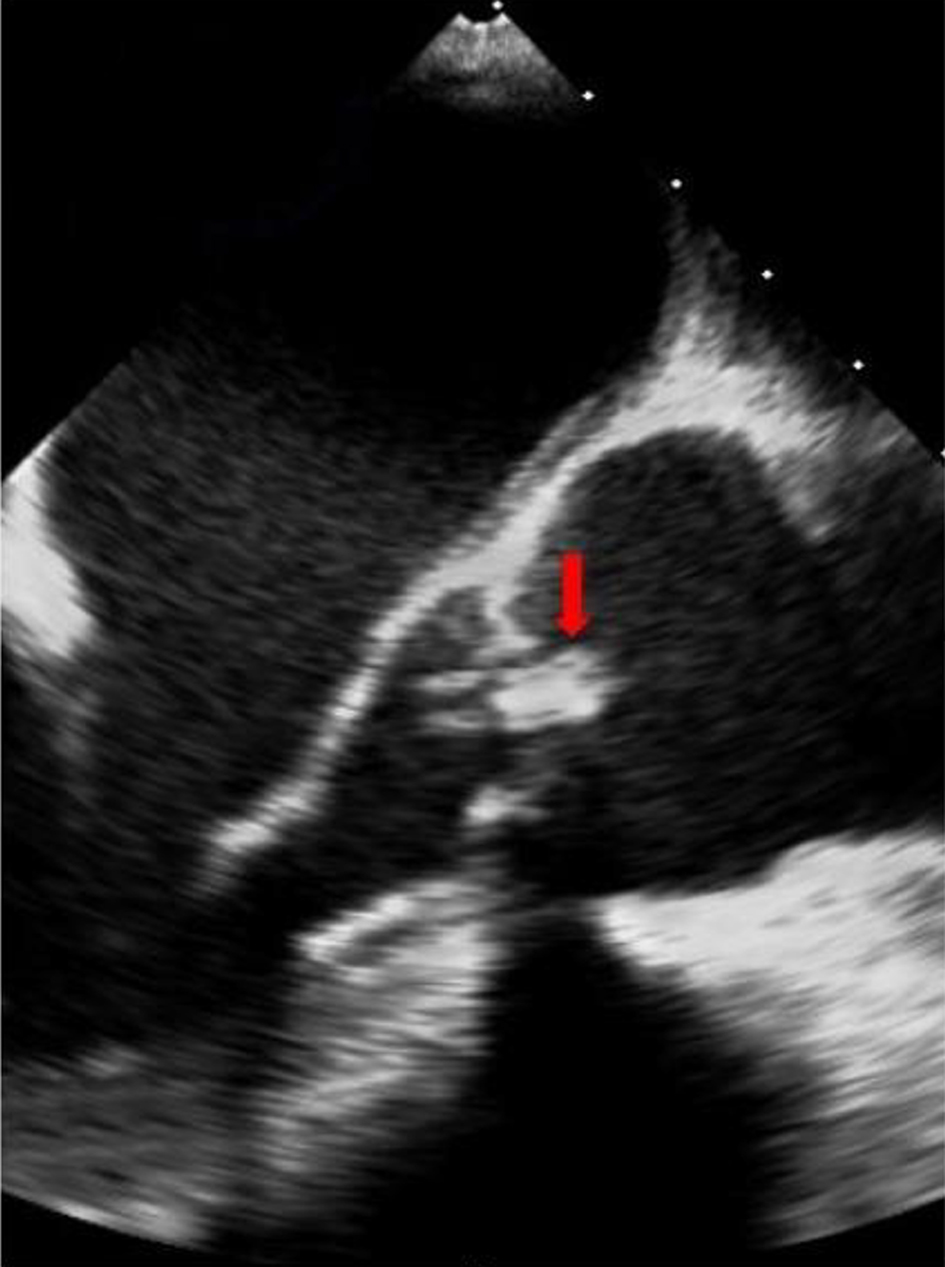
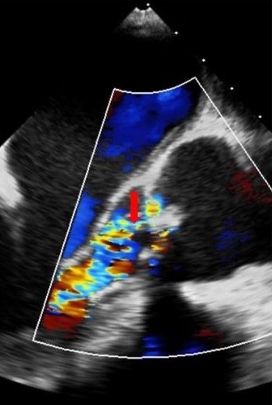
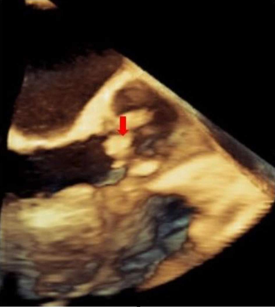
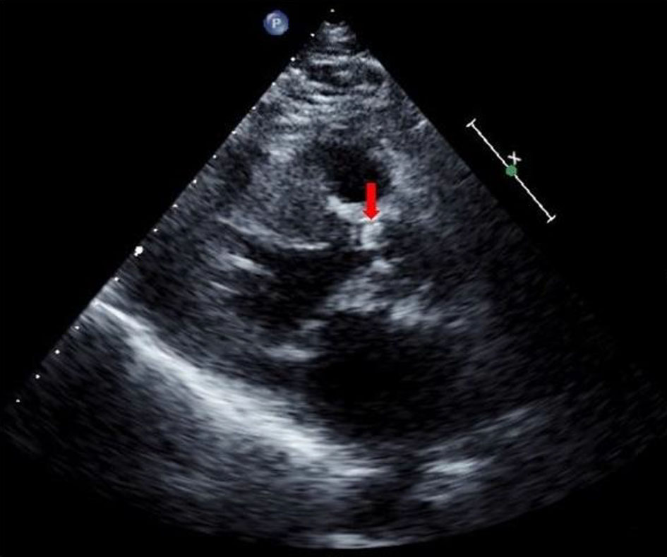
Figure 1. Trans-thoracic paraesternal long-axis view showing a bicuspid, calcified aortic valve without a definitive vegetation image (arrow).
Figure 2.
Figure 2. Transesophageal long-axis view exhibiting an image compatible to a vegetation on a bicuspid aortic valve (arrow).
Figure 3.
Figure 3. Transesophageal long-axis view confirming the presence of double aortic lesion (arrow).
Figure 4.
Figure 4. 3D transesophageal long-axis view showing the vegetation volume and its spacial relationship with the aortic valve (arrow).
Figure 5.
Figure 5. Histology of the native aortic valve (hematoxylin and eosin, ×20 and ×40), showing a thickened and fibrosed valve structure with histological signs of acute endocarditis. Foci of ulceration and fibrinoleukocyte infiltrate with predominance of neutrophils can be observed.