Figures
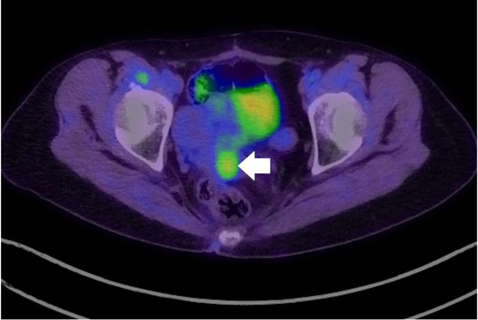
Figure 1. Fluorodeoxyglucose positron emission tomography-computed tomography indicating increased uptake in the cervix (arrow: cervical tumor).
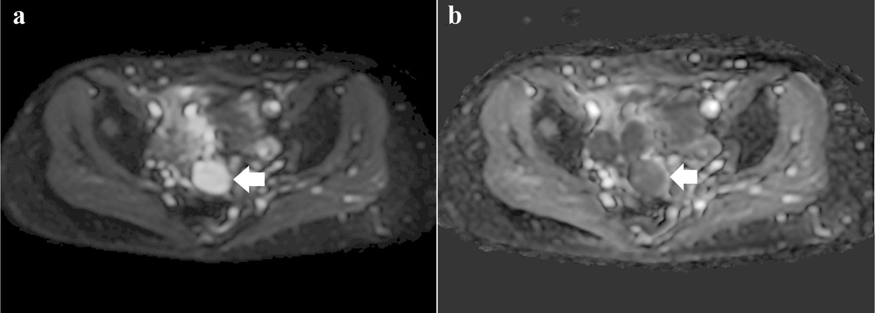
Figure 2. (a) Diffusion-weighted image showing a heterogeneous high-intensity tumor (arrow). (b) Apparent diffusion coefficient image showing a tumor with mild diffusion restriction (arrow).
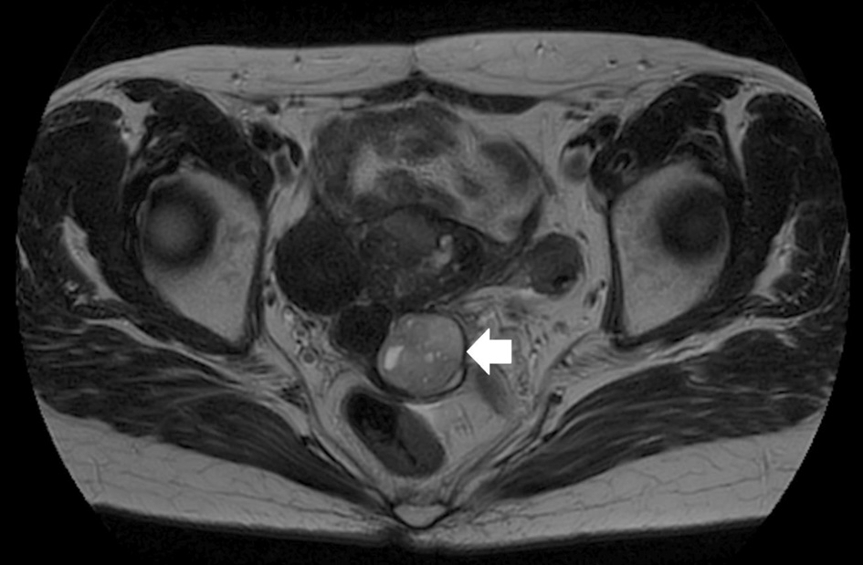
Figure 3. Multiple hyperintense small cysts inside the tumor on T2-weighted imaging (arrow).
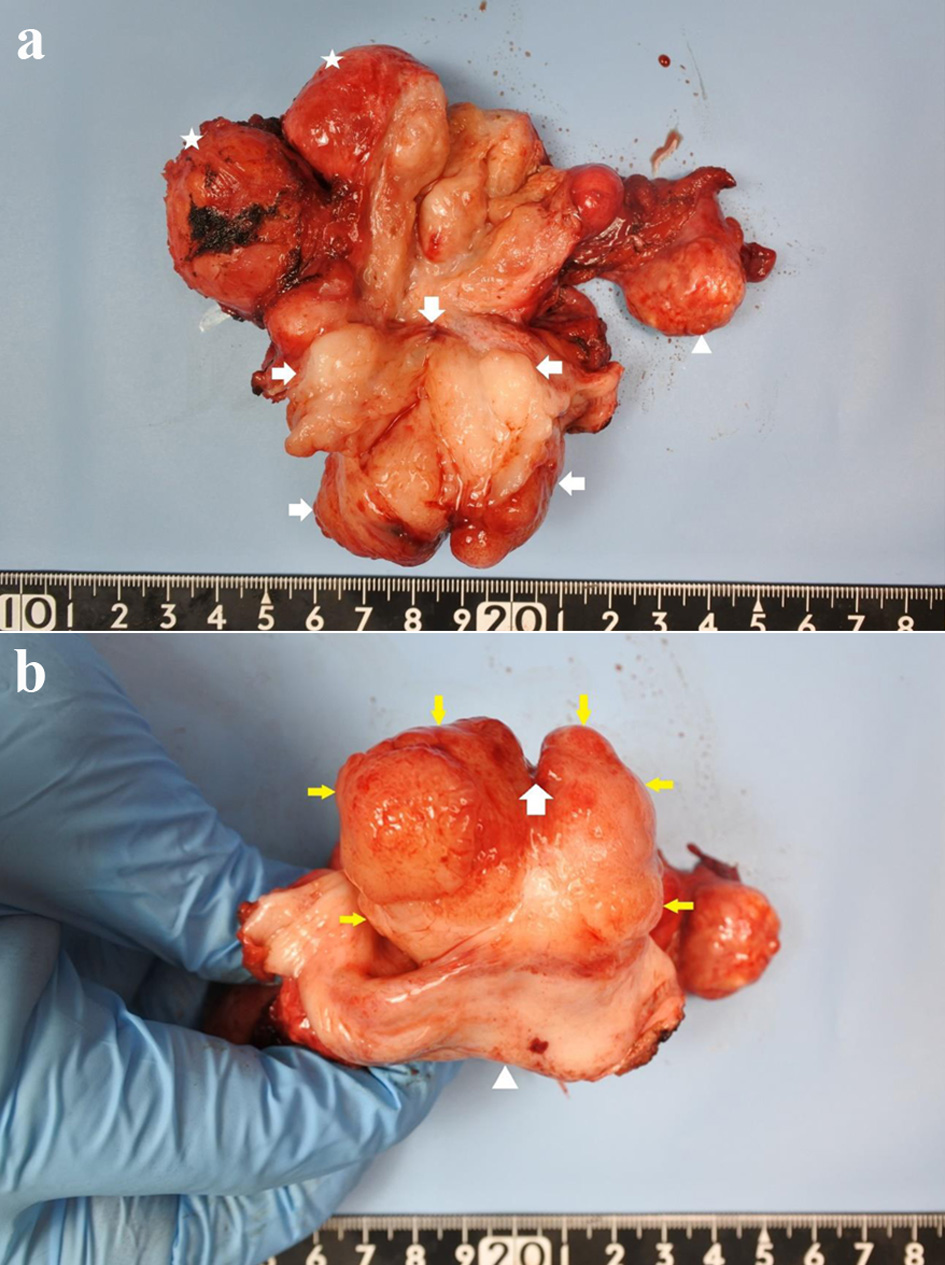
Figure 4. (a) Gross pathological specimen showing a mass occupying the entire cervix (arrowhead: ovary, star: leiomyoma, arrow: cervical tumor). (b) Cervix replaced by the tumor, as seen from the vaginal side (arrow: external os with an incision at 12 o’clock; yellow arrow: cervix; arrowhead: vaginal wall).
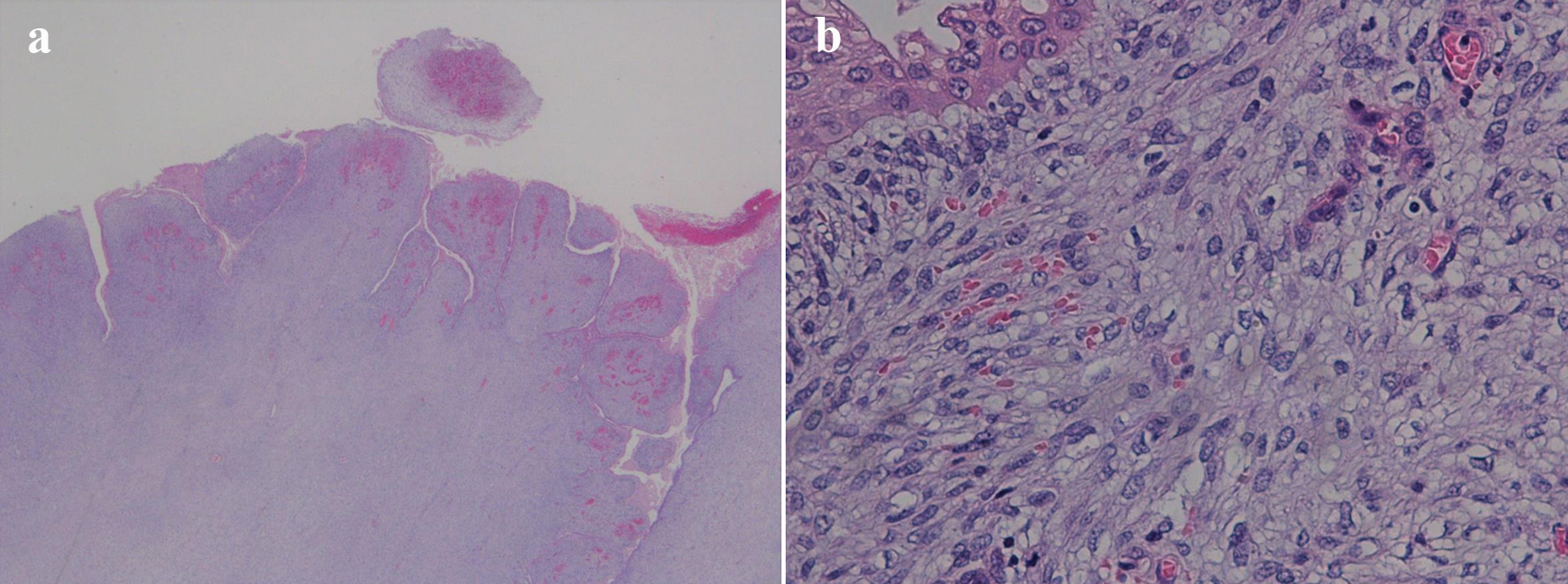
Figure 5. (a) Low-power field showing characteristic leaf-like architecture (hematoxylin and eosin staining, magnification × 10). (b) Papillary stromal fronds lined by benign epithelium accompanied with squamous metaplasia. The stromal cells show mild nucleal atypia and mild pleomorphism (hematoxylin and eosin staining, magnification × 200).
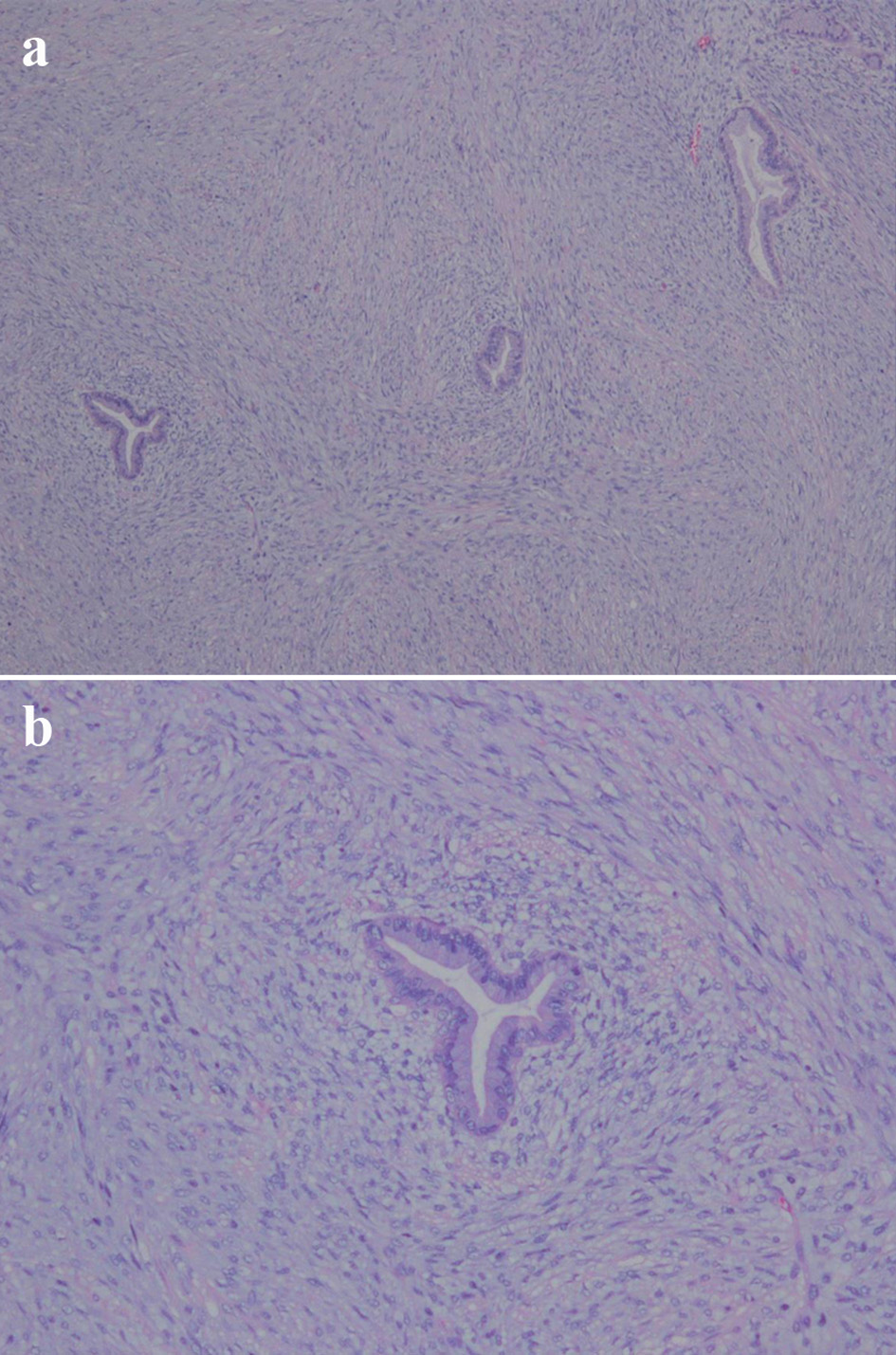
Figure 6. (a) Subepithelial condensation of the stroma; periglandular cuffing (hematoxylin and eosin staining, magnification × 40). (b) Hypercellular stroma and high cellularity beneath the epithelium (hematoxylin and eosin staining, magnification × 100).
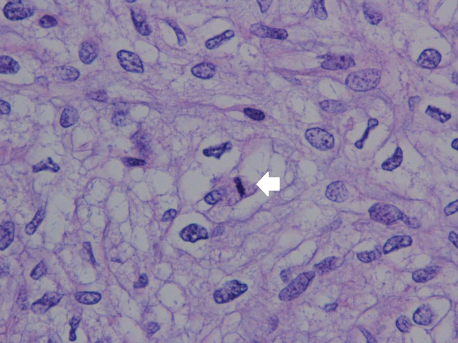
Figure 7. Stromal atypia with mitosis in the stromal element (arrow) (hematoxylin and eosin staining, magnification × 400).
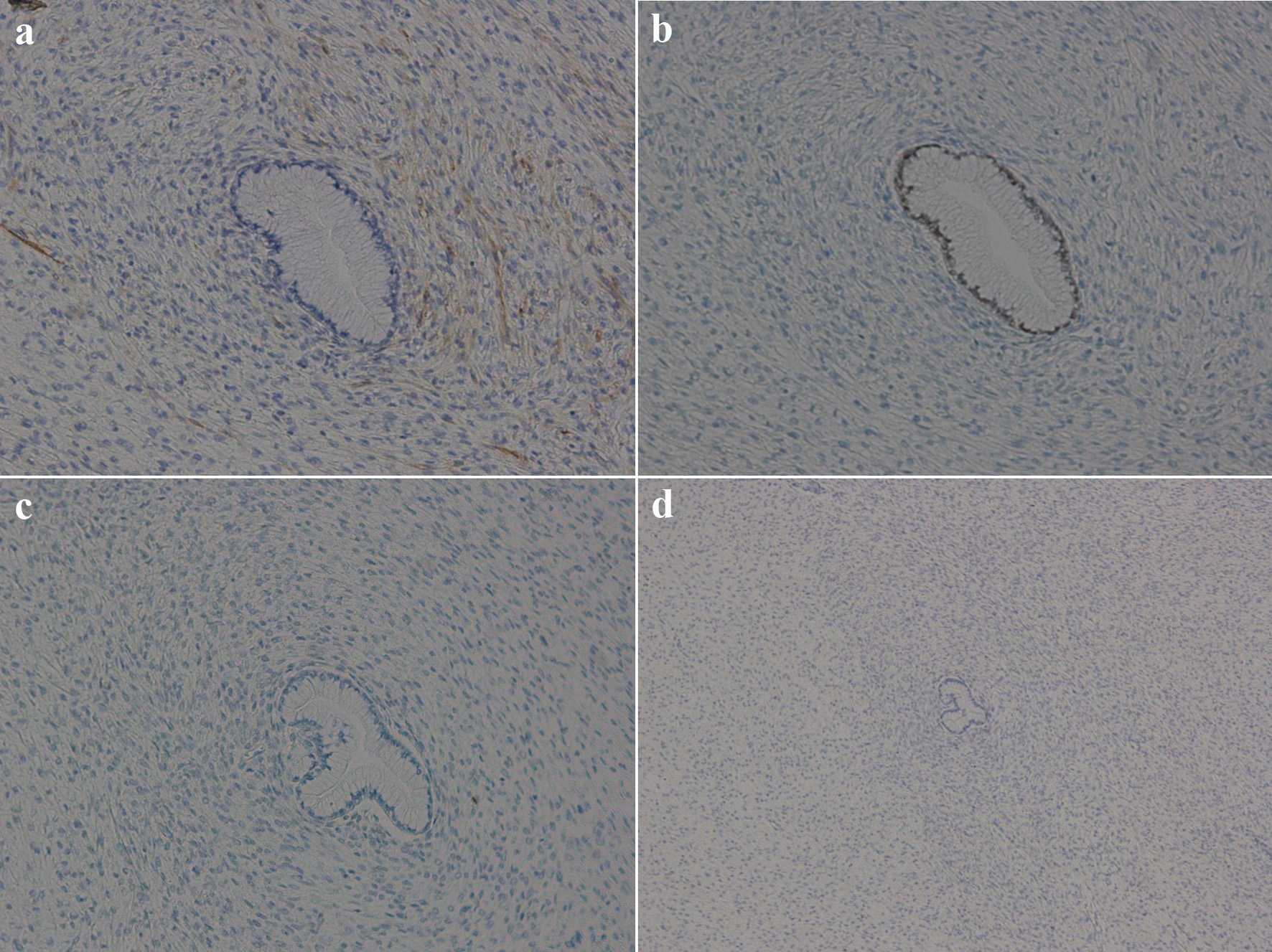
Figure 8. Immunohistochemical findings of the tumor. (a) Stromal cell cytoplasm positive for CD10 (magnification, ×100); (b) glandular epithelia nuclei positive but stromal cell nuclei negative for estrogen receptors (magnification, ×100); (c) both glandular epithelia nuclei and stromal cell nuclei negative for progesterone receptors (magnification, ×100); (d) stromal cell showing wild-type pattern for P53 (magnification, ×40).