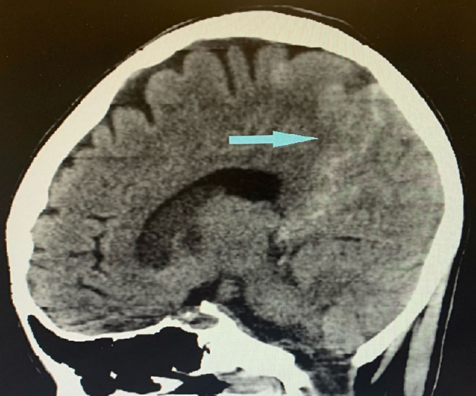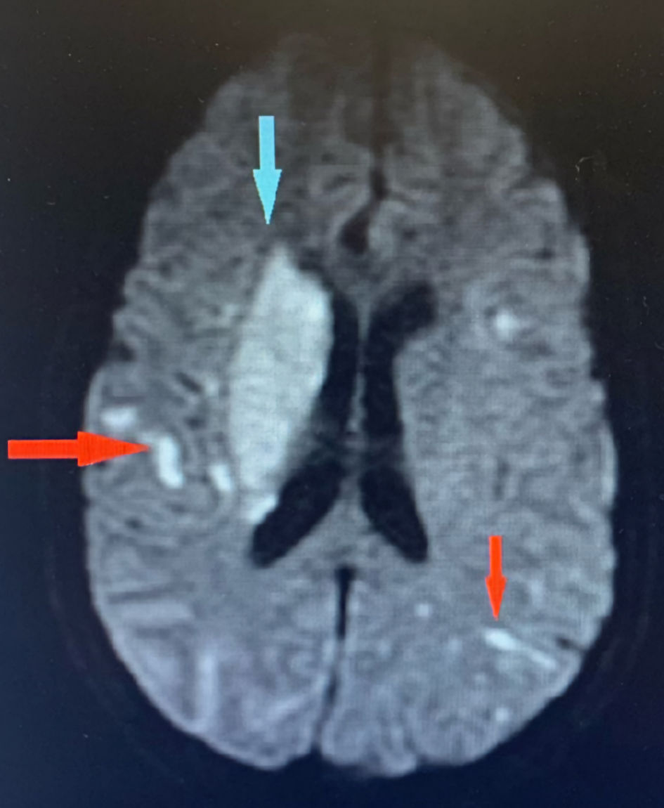
Figure 1. Brain MRI showing innumerable acute to subacute embolic infarcts in both cerebral hemispheres (red arrows). The largest infarct involves the right lentiform nucleus and corona radiate (blue arrow). MRI: magnetic resonance imaging.
| Journal of Medical Cases, ISSN 1923-4155 print, 1923-4163 online, Open Access |
| Article copyright, the authors; Journal compilation copyright, J Med Cases and Elmer Press Inc |
| Journal website https://www.journalmc.org |
Case Report
Volume 13, Number 7, July 2022, pages 349-353
A Unique Case of Tri-Valvular Serratia marcescens Infective Endocarditis Complicated by Innumerable Emboli
Figures