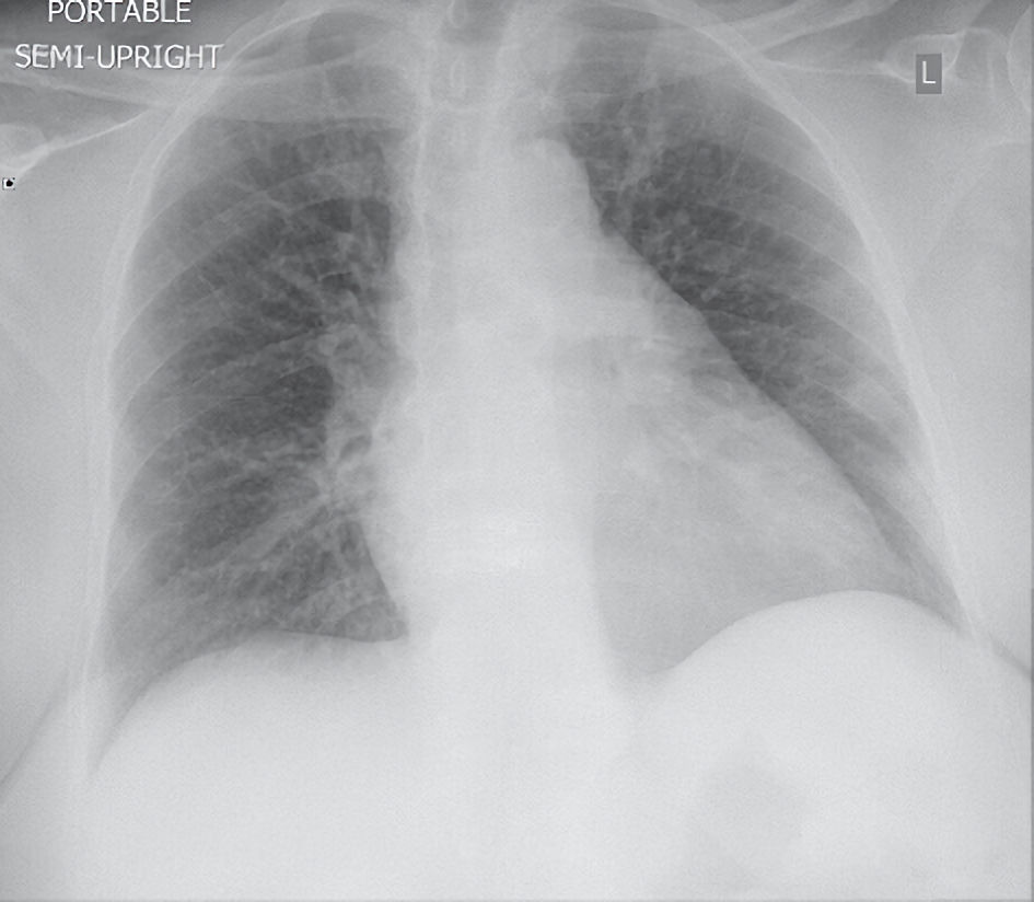
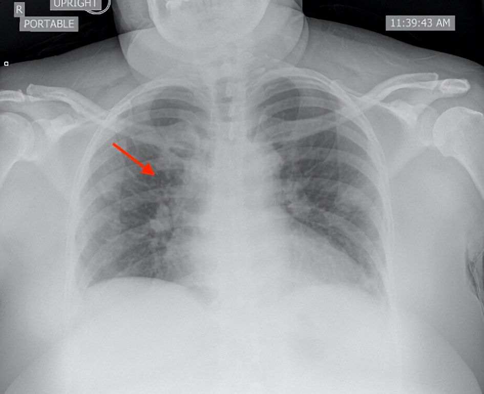
Figure 1. Posteroanterior chest X-ray showed no infiltrate, effusion, or pneumothorax.
| Journal of Medical Cases, ISSN 1923-4155 print, 1923-4163 online, Open Access |
| Article copyright, the authors; Journal compilation copyright, J Med Cases and Elmer Press Inc |
| Journal website https://www.journalmc.org |
Case Report
Volume 13, Number 8, August 2022, pages 380-385
Functionality of Monoclonal Antibody Therapy in SARS-CoV-2
Figures
Table
| Case 1 | Case 2 | Case 3 | Reference values | |
|---|---|---|---|---|
| Blood urea nitrogen (mg/dL) | 12 | 65 | 7 | 6 - 24 |
| Serum creatinine (mg/dL) | 0.6 | 2.6 | 0.8 | 0.5 - 1 |
| White blood cell counts (× 103/µL) | 8.7 | 8.6 | 2.5 | 4.4 - 11 |
| Hemoglobin (g/dL) | 14 | 8.2 | 12.5 | 12 - 15.5 |
| Platelets (× 103/µL) | 319 | 41 | 111 | 150 - 450 |
| Lactate dehydrogenase (U/L) | 484 | 420 | 426 | 122 - 222 |
| C-reactive protein (mg/dL) | 22.2 | 11.8 | 18.3 | 0.0 - 0.8 |
| Ferritin (ng/mL) | 696 | 371 | 838 | 11 - 307 |
| D-dimer (ng/mL) | 6,059 | 7,771 | 658 | 0 - 500 |