Journal of Medical Cases, ISSN 1923-4155 print, 1923-4163 online, Open Access
Article copyright, the authors; Journal compilation copyright, J Med Cases and Elmer Press Inc
Journal website http://www.journalmc.org

Case Report

Volume 2, Number 6, December 2011, pages 275-278


Hydrocele and Unexpected Diagnosis: Malignant Mesothelioma of Tunica Vaginalis Testis

Figures

Figure 1.
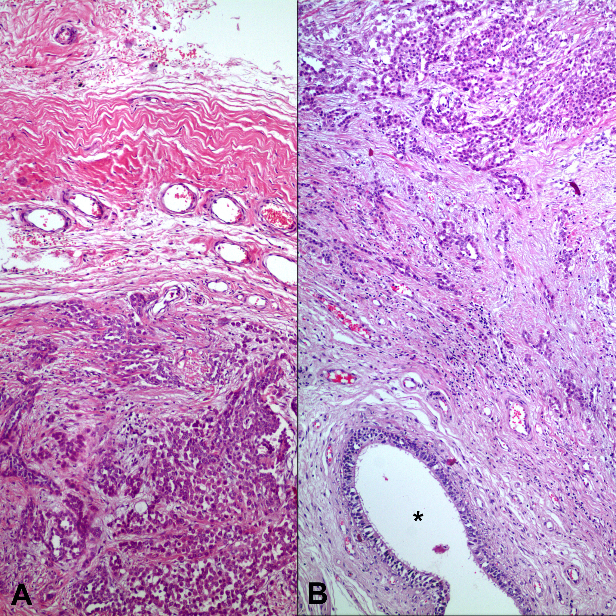
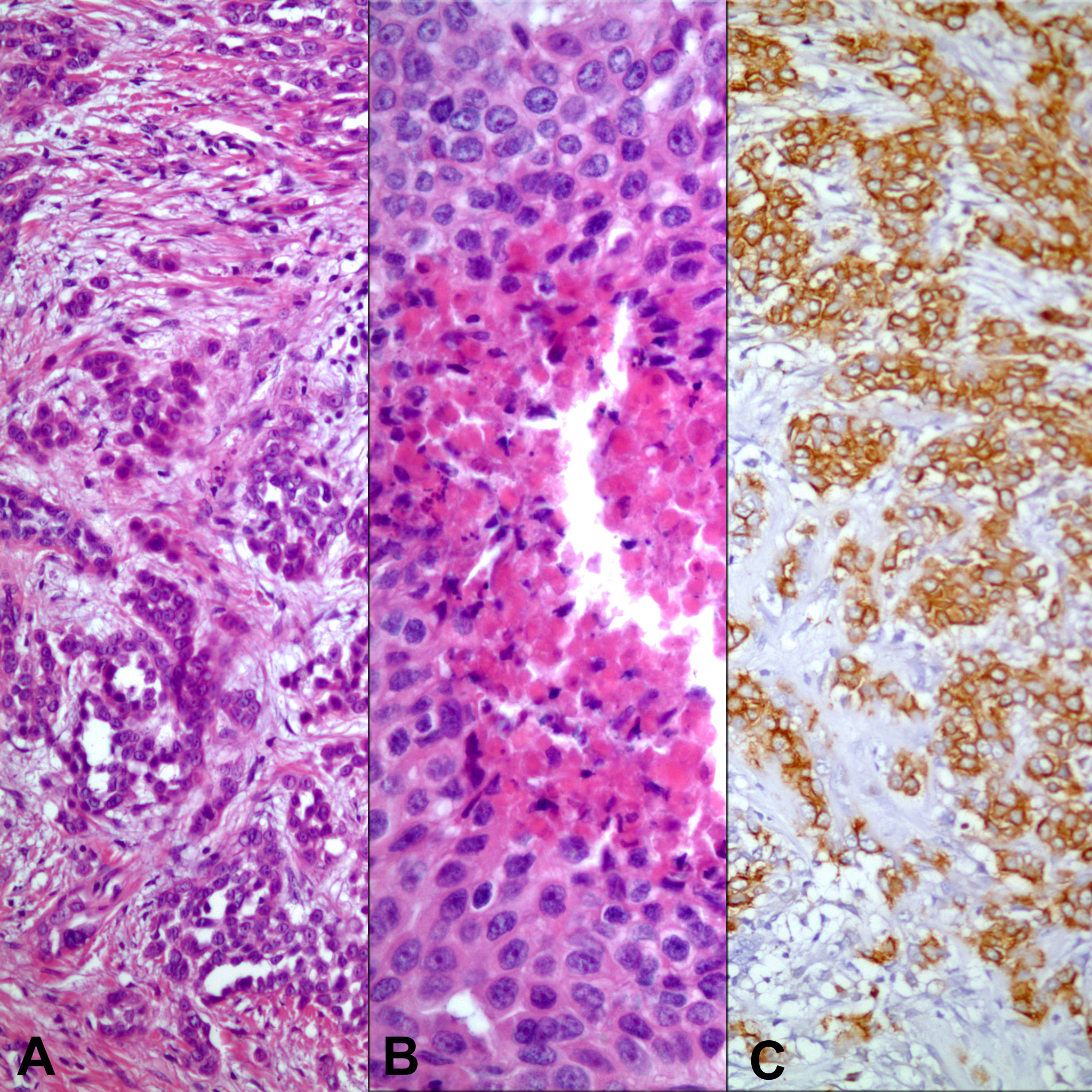
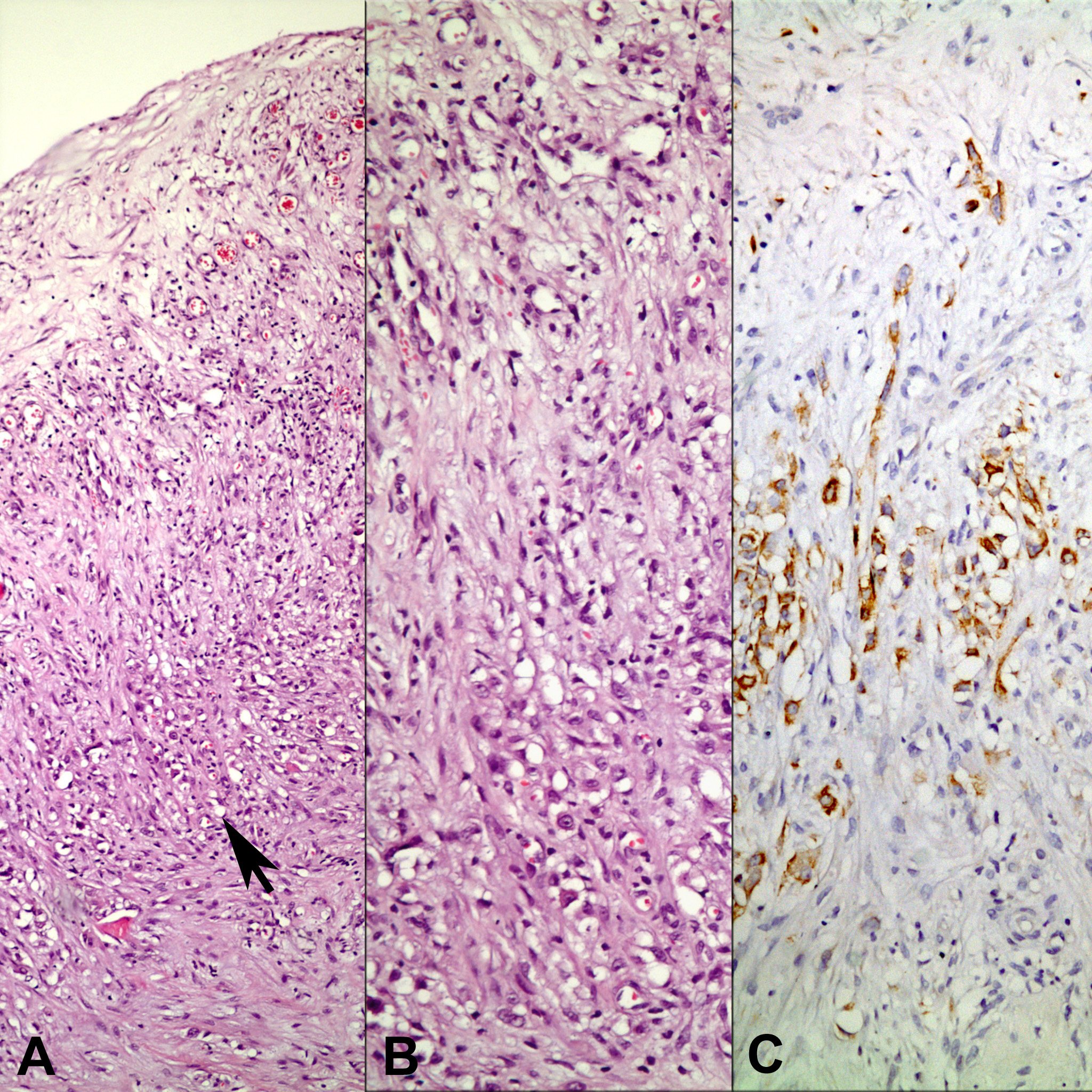
Figure 1. Biopsy from tunica vaginalis showing mesothelial cell proliferation (arrow). Panel B and C represent the area pointed by the arrow (A: H-E x 40; B: H-E x 200; C: Immunohistochemistry, anti-calretinin Ab x 200).
Figure 2.
Figure 2. Macroscopical appearance of the tumor (A: Before fixation; B: After fixation).
Figure 3.
Figure 3. Tumor infiltrating paratesticular tissues. *shows an epidydimal ductus (A: H-E x 100; B: H-E x 40).
Figure 4.
Figure 4. Neoplasm showing tubular (panel A) and solid patterns with necrosis (panel B). Neoplastic cells diffusely express a mesothelial marker, HBME-1 (panel C) (A: H-E x 200; B: H-E x400; C: Immunohistochemistry, anti- HBME-1 Ab x 200).
Figure 5.
Figure 5. One of the lymph nodes with metastasis of mesothelioma (A: H-E x 200; B: Immunohistochemistry, anti-calretinin Ab x 200).