Journal of Medical Cases, ISSN 1923-4155 print, 1923-4163 online, Open Access
Article copyright, the authors; Journal compilation copyright, J Med Cases and Elmer Press Inc
Journal website https://www.journalmc.org

Case Report

Volume 14, Number 1, January 2023, pages 1-6


Clinically Isolated Syndrome and Frontal Lobe Arteriovenous Malformation Presenting With Behavior Issues

Figures

Figure 1.
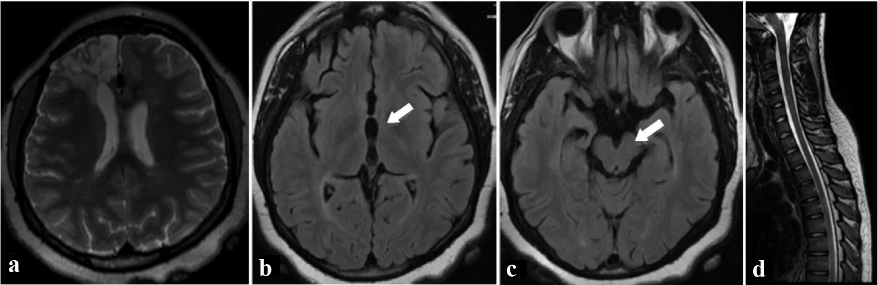
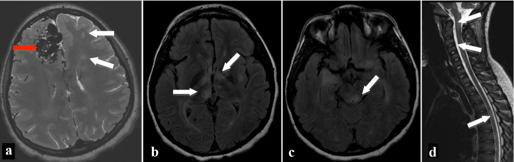
Figure 1. MRI of brain on axial T2-weighted image (a) shows multiple flow voids in the right frontal lobe (red arrow) consistent with arteriovenous malformation as well as multiple hyperintense T2 signal areas (white arrows) in the white matter bilaterally. Fluid-attenuated inversion recovery axial images show hyperintensities in the bilateral thalami (b), left globus pallidus, left putamen, bilateral cerebral white matter, and brainstem (c). MRI of the spine shows patchy hyperintense signal areas (arrows) in the brainstem, cervical spinal cord, and thoracic spinal cord on T2-weighted sagittal image (d). MRI: magnetic resonance imaging.
Figure 2.
Figure 2. Two years after initial presentation, T2-weighted axial (a) MRI image shows resection bed of removed arteriovenous malformation from right frontal lobe. Fluid-attenuated inversion recovery axial (b, c) MRI images of the brain show resolution of white matter lesions in the thalamus (a) and brainstem (b). There was a decrease in the number and size of other lesions during the interval, and no new lesions were found. T2-weighted sagittal (d) MRI image of spine shows interval improvement in hyperintense T2 signal areas in the spinal cord. MRI: magnetic resonance imaging.

Tables

Table 1. Results of Lumbar Puncture
 
Cerebrospinal fluid findingsValue
aElevated or positive results. IgG: immunoglobulin G.
White blood cell count (µL)9
Red blood cell count (µL)1675
Neutrophils (%)8
Lymphocytes (%)79
Monocytes (%)13
Glucose (mg/dL)63
IgGa (mg/dL)10.30
Proteina (mg/dL)105
Oligoclonal bandingaPositive
IgG nephelometer (mg/dL)994
IgG/albumina (%)100.0
IgG indexa3.18
IgG synthesisa (mg/day)40.36
Angiotensin-converting enzyme (U/L)< 5


Table 2. Laboratory Evaluation of Acute Central Nervous System Demyelination in Children: CSF and Serum Studies and Their Corresponding Disease Processes
 
ARSA-A: arylsulfatase A; CSF: cerebrospinal fluid; GFAP: glial fibrillary acidic protein; NMOsd: neuromyelitis optica spectrum disorder; POMS: pediatric onset multiple sclerosis; VLCFA: very long chain fatty acid.
CSF
  Angiotensin-converting enzymeNeurosarcoidosis
  Oligoclonal bandingPOMS
Serum
  NMO-IgG (aquaporin-4)NMOsd
  West Nile virus IgM antibodyWest Nile virus
  B12 levelB12 deficiency
  Folate levelFolate deficiency
  ARSA-A enzyme levelMetachromatic leukodystrophy
  Double-stranded DNA antibodiesSystemic lupus erythematosus
  GFAP assayAlexander disease
  VLCFA panel (males)X-linked adrenoleukodystrophy
  ABCD1 gene mutation analysis (females)X-linked adrenoleukodystrophy