Journal of Medical Cases, ISSN 1923-4155 print, 1923-4163 online, Open Access
Article copyright, the authors; Journal compilation copyright, J Med Cases and Elmer Press Inc
Journal website https://www.journalmc.org

Case Report

Volume 13, Number 10, October 2022, pages 504-508


The Management of Traumatic Abdominal Wall Flank Hernia Along the Spigelian Aponeurosis Using Component Separation, Synthetic, and Biological Mesh

Figures

Figure 1.
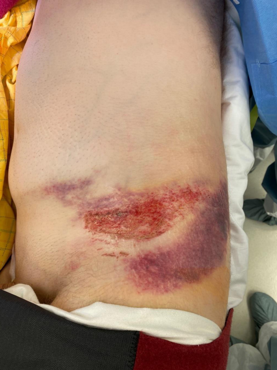
Figure 1. Ecchymosis noted on the patient’s left flank and hip.
Figure 2.
Figure 2. Sagittal, transverse, and coronal computed tomography of abdomen showing disruption of left abdominal wall musculature from iliac crest and retraction of the muscle noted by a white arrow in the three figure segments.
Figure 3.
Figure 3. (a) Biological mesh inlay bridging transversus abdominis to iliac crest to recreate visceral sac. (b) Ventralight™ mesh underlay secured to internal oblique and iliac crest. (c) Counter incision visualized for access to retracted abdominal wall musculature. (d) Re-approximation of internal oblique muscle over mesh repair.