Figures
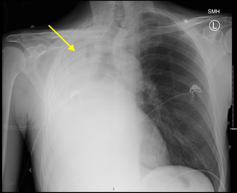
Figure 1. Chest X-ray with arrow depicting complete opacification of the right hemithorax with mediastinal shift to the right. Mild irregularity of the right mainstem bronchus.
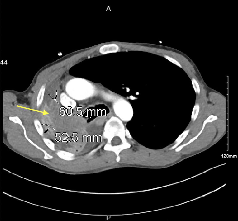
Figure 2. Contrast-enhanced CT image with yellow arrow depicting ill-defined hypoattenuation within the right upper lobe ascending into the right mainstem bronchus and bronchus intermedius measuring 6.1 × 5.3 cm. CT: computed tomography.
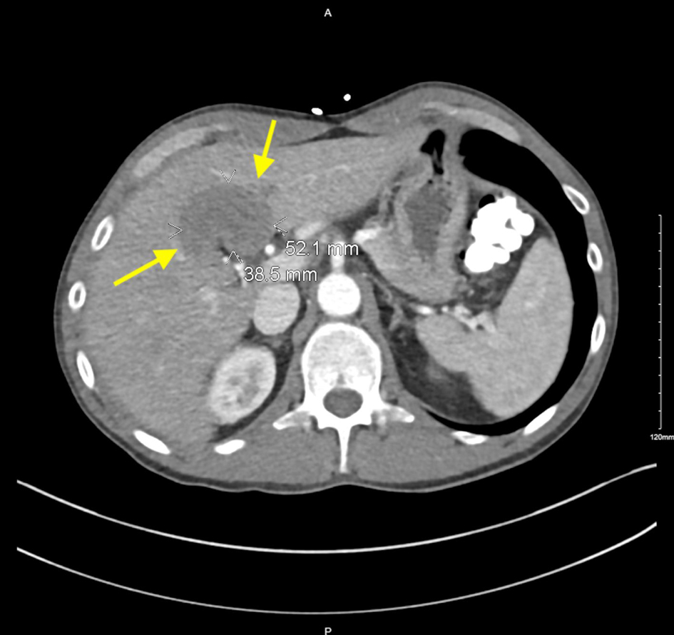
Figure 3. Contrast-enhanced CT image with yellow arrows pointing to hypodense lesion in the liver measuring 5.2 × 3.8 cm. CT: computed tomography.
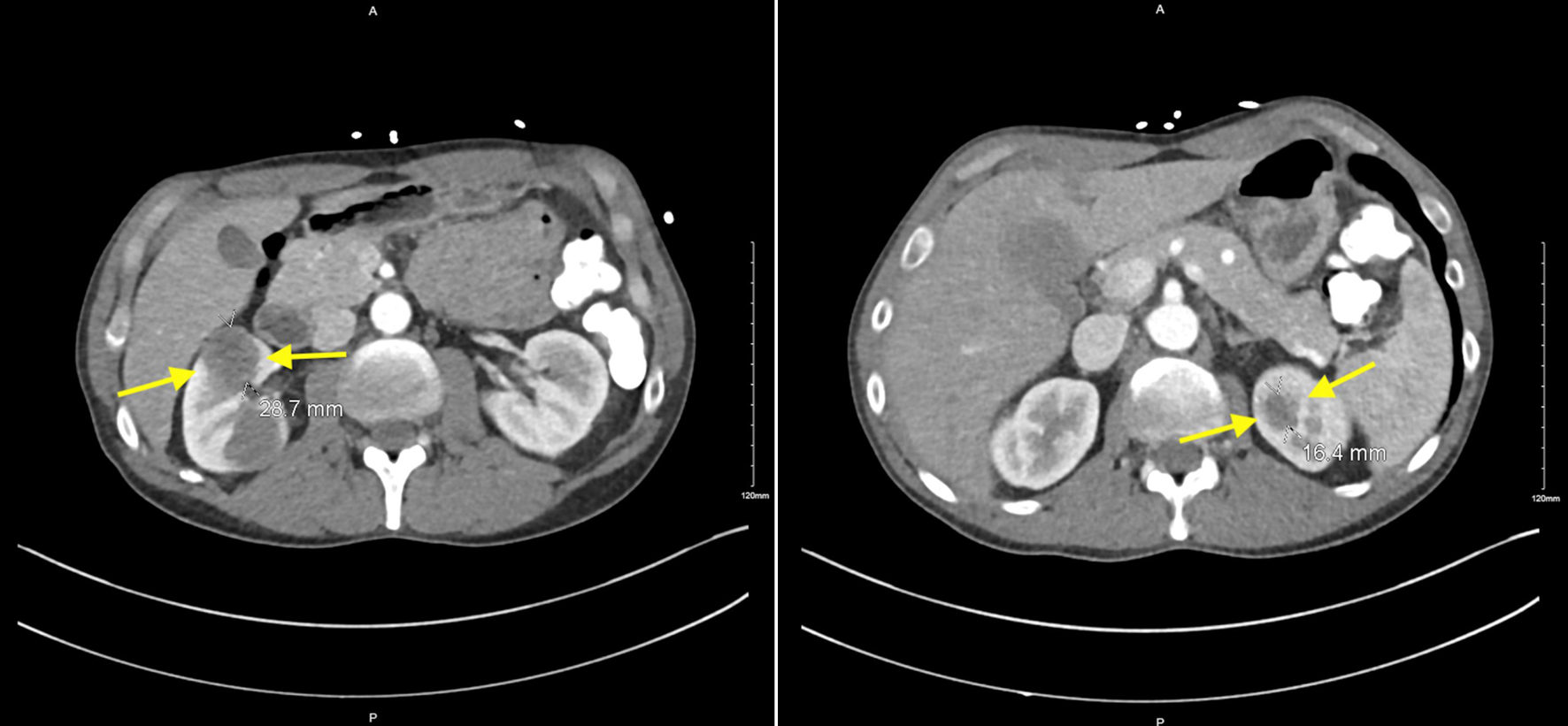
Figure 4. Contrast-enhanced CT imaging with yellow arrows depicting multiple bilateral renal hypodensities. Right anterior interpolar region measuring 2.9 cm and within the left upper pole measuring 1.6 cm. CT: computed tomography.
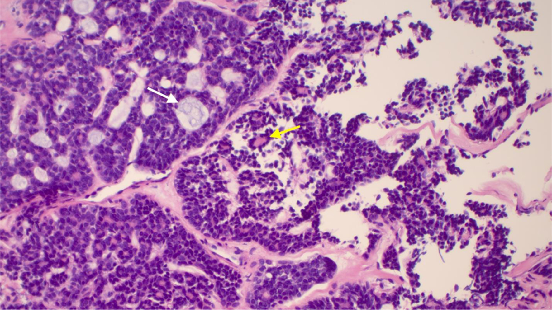
Figure 5. Right mainstem bronchus with adenoid cystic carcinoma in hematoxylin and eosin stain. White arrow depicts cribriform island containing basophilic mucin surrounded by myoepithelial cells. Yellow arrow depicts epithelial island with luminal cells.
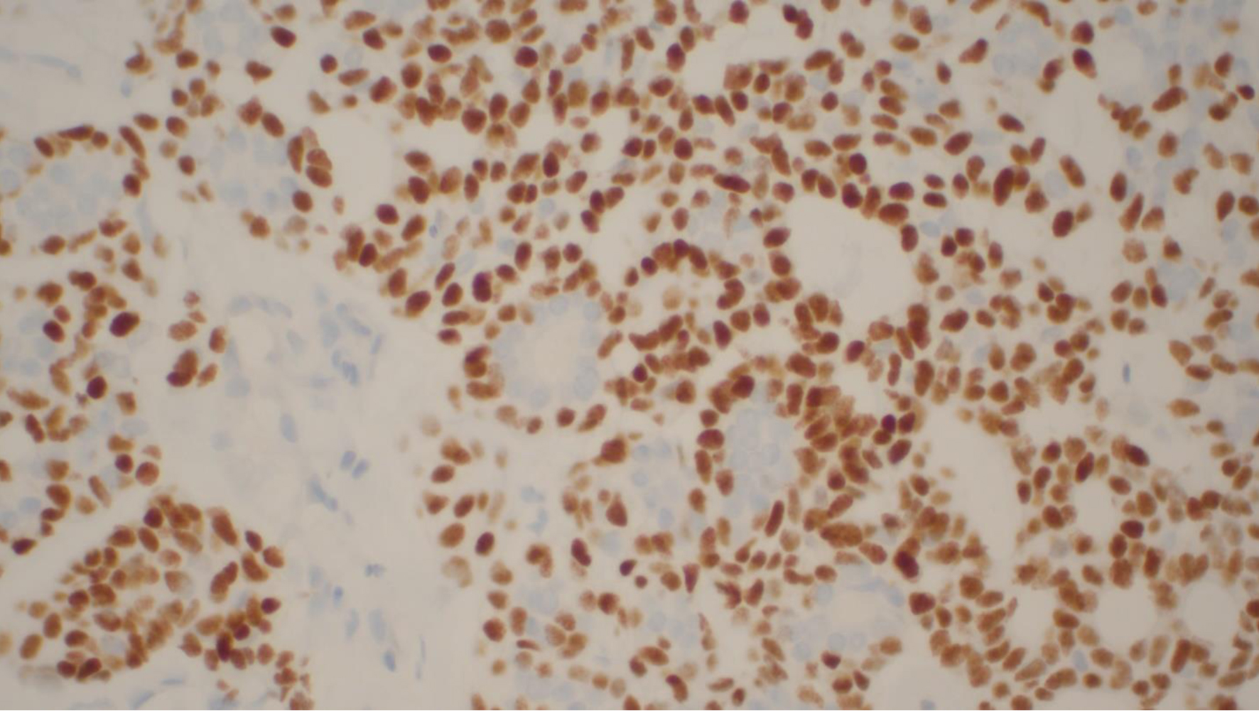
Figure 6. Adenoid cystic carcinoma with immunoperoxidase p40 staining nuclei of myoepithelial cells. Luminal epithelial cells are unstained.
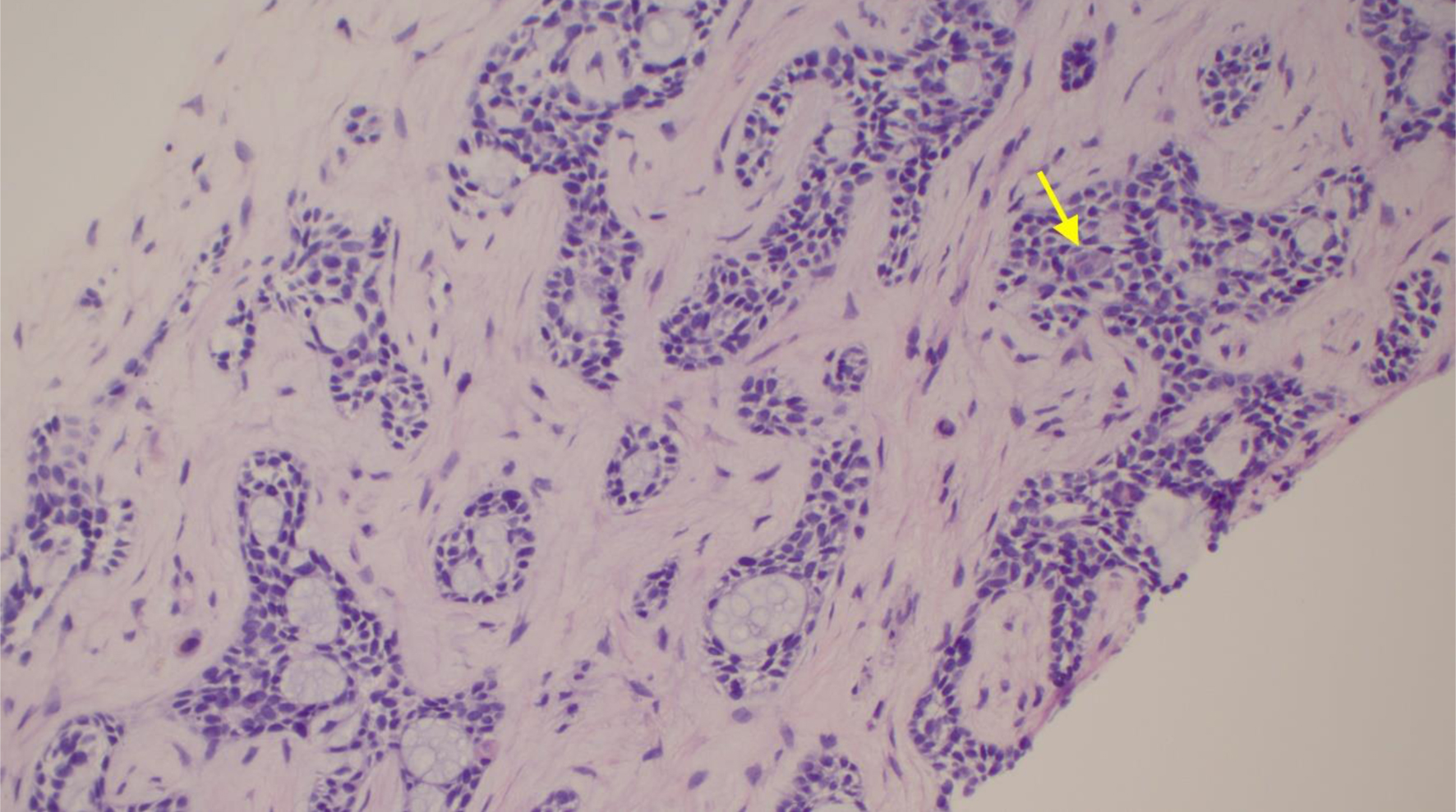
Figure 7. Section of liver core in hematoxylin and eosin stain showing identical morphology. Yellow arrow shows epithelial cells. Bluish mucin surrounding myoepithelial cells.