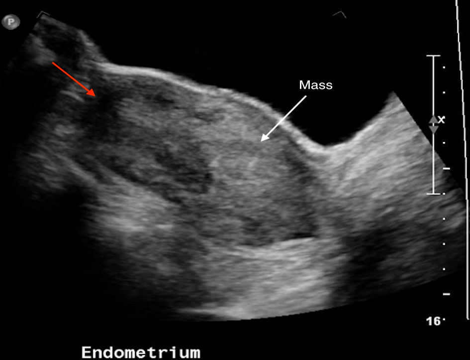
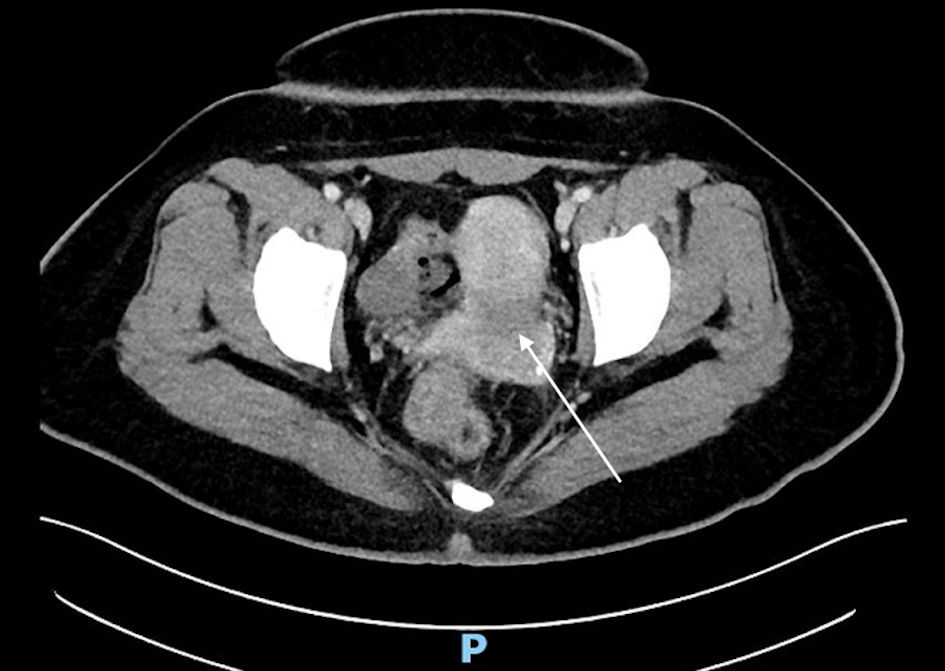
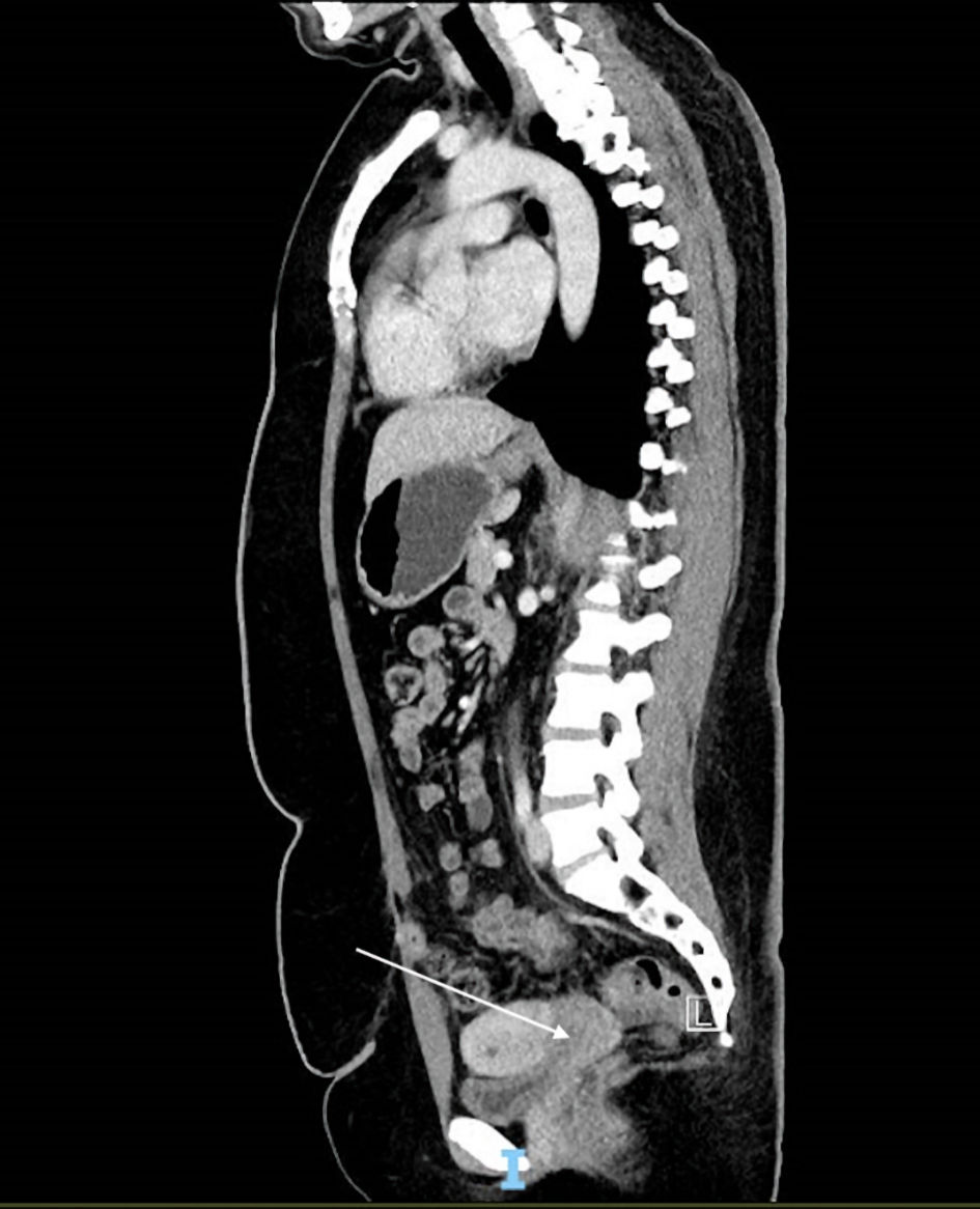
Figure 1. Ultrasound image with white arrow pointing to poorly defined mass within endometrial cavity. Red arrow indicates area where uterine fundus should be but was not well seen.
| Journal of Medical Cases, ISSN 1923-4155 print, 1923-4163 online, Open Access |
| Article copyright, the authors; Journal compilation copyright, J Med Cases and Elmer Press Inc |
| Journal website https://www.journalmc.org |
Case Report
Volume 14, Number 1, January 2023, pages 7-12
Uterine Inversion Secondary to Endometrial Carcinoma
Figures
Tables
| Stage 1 | Incomplete inversion with the uterine fundus remaining within the cavity |
| Stage 2 | Complete inversion of the uterine fundus through the fibromuscular ring of the cervix |
| Stage 3 | Total inversion with the fundus protruding through the vulva |
| Stage 4 | The vagina is also involved and inverted through the vulva along with the uterus |
| Method | Route | Description |
|---|---|---|
| Johnson’s maneuver | Conservative (vaginal) | Manual reduction by placing a hand inside the vagina and pushing the fundus along the long axis of the vagina towards the umbilicus. |
| O’Sullivan’s method | Conservative (vaginal) | Infusing warm saline into the vagina, therefore creating a water seal with the operator’s hand and the vulva. |
| Ogueh and Ayida technique (modified O’Sullivan’s method) | Conservative (vaginal) | An intravenous fluid tubing is connected to the silicon cup used in vacuum deliveries after the cup is placed in the vagina, creating a water seal when fluid is infused. |
| Huntington’s method | Surgical (abdominal) | Sequential clamping and upward traction on the round ligaments using Allis/Babcock clamps starting from approximately 2 cm deep in the cup formed by the inversion. If the round ligaments are unable to be identified, the myometrium may be clamped instead. |
| Haultain procedure | Surgical (abdominal) | Incising the posterior surface of the uterus to bisect the constriction ring. This may then allow the inversion to be reduced manually or via Huntington’s method. |
| Spinelli’s method | Surgical (vaginal) | Transecting the cervix anteriorly to bisect the constriction ring. This will be followed by manual reduction. |
| Kustner’s method | Surgical (vaginal) | Transecting the cervix posteriorly to bisect the constriction ring. This will be followed by manual reduction. |