Journal of Medical Cases, ISSN 1923-4155 print, 1923-4163 online, Open Access
Article copyright, the authors; Journal compilation copyright, J Med Cases and Elmer Press Inc
Journal website https://www.journalmc.org

Case Report

Volume 14, Number 1, January 2023, pages 13-18


Breast to Brain: A Case Report and Literature Review of Leptomeningeal Carcinomatosis

Figures

Figure 1.
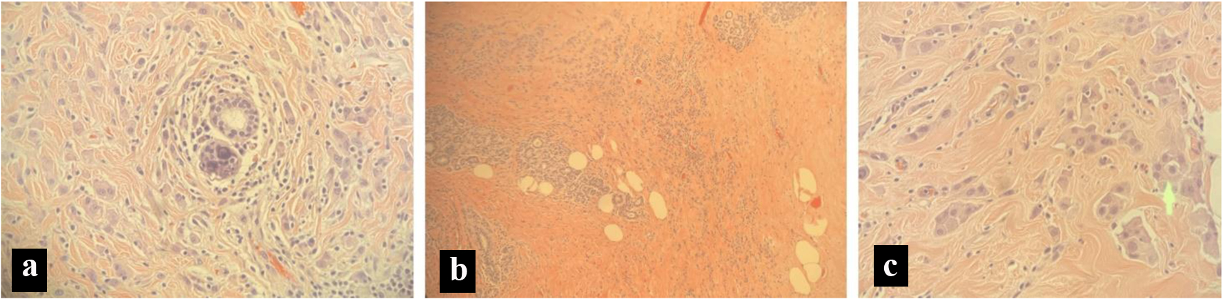
Figure 1. Pathology slides from a breast biopsy. (a) Micro calculi in a ductal pattern from left breast tissue. (b) Stromal sclerosis and infiltrating lobular cancer from left breast tissue. (c) Cells in a single file line, indicating lobular carcinoma from left breast tissue.
Figure 2.
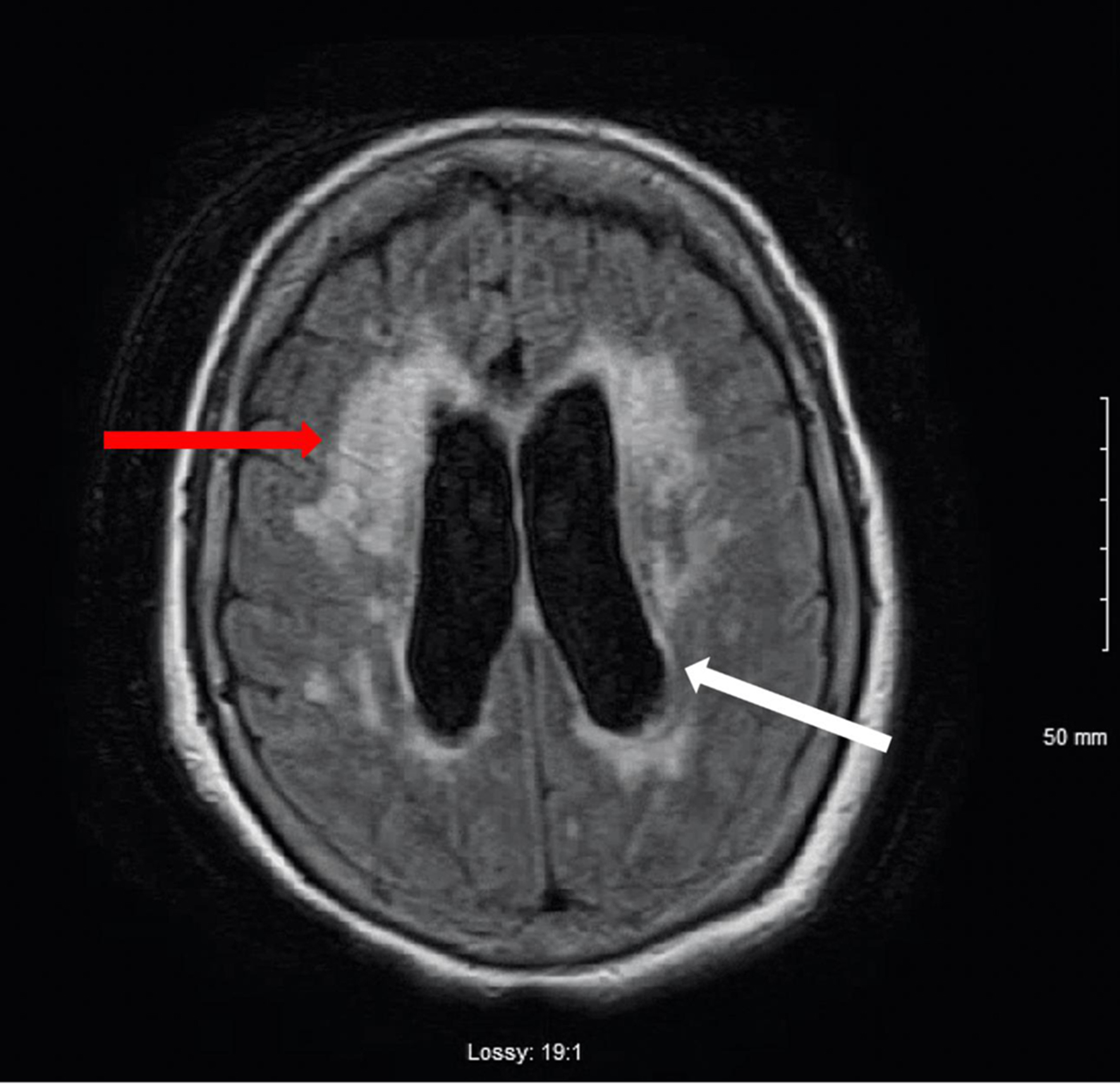
Figure 2. T1-weighted MRI image of the brain. Mild to moderate hydrocephalus (white arrow). T2 hyperintensity consistent with transependymal spread of CSF (red arrow). There is no evidence of mass. There is no abnormal gadolinium enhancement. CSF: cerebrospinal fluid; MRI: magnetic resonance imaging.
Figure 3.
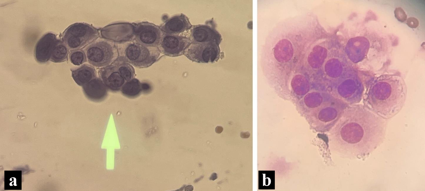
Figure 3. Pathology slides form the cerebrospinal fluid. (a) Enlarged nucleoli with cytoplasmic vacuoles on cerebrospinal fluid cytology (May Grunwald-Giemsa stain). (b) Wright stain indicating a nest of tumors with increased cytoplasm, non-spindled from cerebrospinal fluid cytology.
Figure 4.
Figure 4. Algorithm for the correct diagnostic approach to leptomeningeal carcinomatosis.

Tables

Table 1. Demographic Details and Diagnostic Modalities for Leptomeningeal Carcinomatosis (Review of Literature)
 
ReferencesAge (years)SexERPRHER-2Duration from breast cancer diagnosis to LC diagnosis (years)Imaging
F: female; M: male; ER: estrogen receptor; PR: progesterone receptor; HER-2: human epidermal growth factor receptor 2; LC: leptomeningeal carcinomatosis; MRI: magnetic resonance imaging; CT: computed tomography; PET: positron emission tomography; CSF: cerebrospinal fluid; -: not mentioned.
[3]65FPositivePositiveNegative18PET, MRI
[4]71FPositivePositiveNegative0MRI
[5]67F---2MRI
[6]66FNegativeNegativePositive2MRI
[7]77F-----
[8]66FPositive-Negative0MRI


Table 2. Significant Laboratory Results From the Cerebrospinal Fluid
 
Spinal fluidPatient value (units)Reference range
µL: microliter; dL: deciliter; mg: milligram; RBCs: red blood cells; WBCs: white blood cells; CSF: cerebrospinal fluid; VDRL: Venereal Disease Research Laboratory.
Glucose, CSF91 mg/dL40 - 70 mg/dL
Protein, CSF365 mg/dL15 - 45 mg/dL
RBC, CSF5 µL0 µL
WBC, CSF25 µL0 µL
CSF VDRLNon-reactive
Volume, CSF2.75 mL