Journal of Medical Cases, ISSN 1923-4155 print, 1923-4163 online, Open Access
Article copyright, the authors; Journal compilation copyright, J Med Cases and Elmer Press Inc
Journal website https://www.journalmc.org

Case Report

Volume 14, Number 1, January 2023, pages 25-30


A Case of Antisynthetase Syndrome Initially Presented With Interstitial Lung Disease Mimicking COVID-19

Figures

Figure 1.
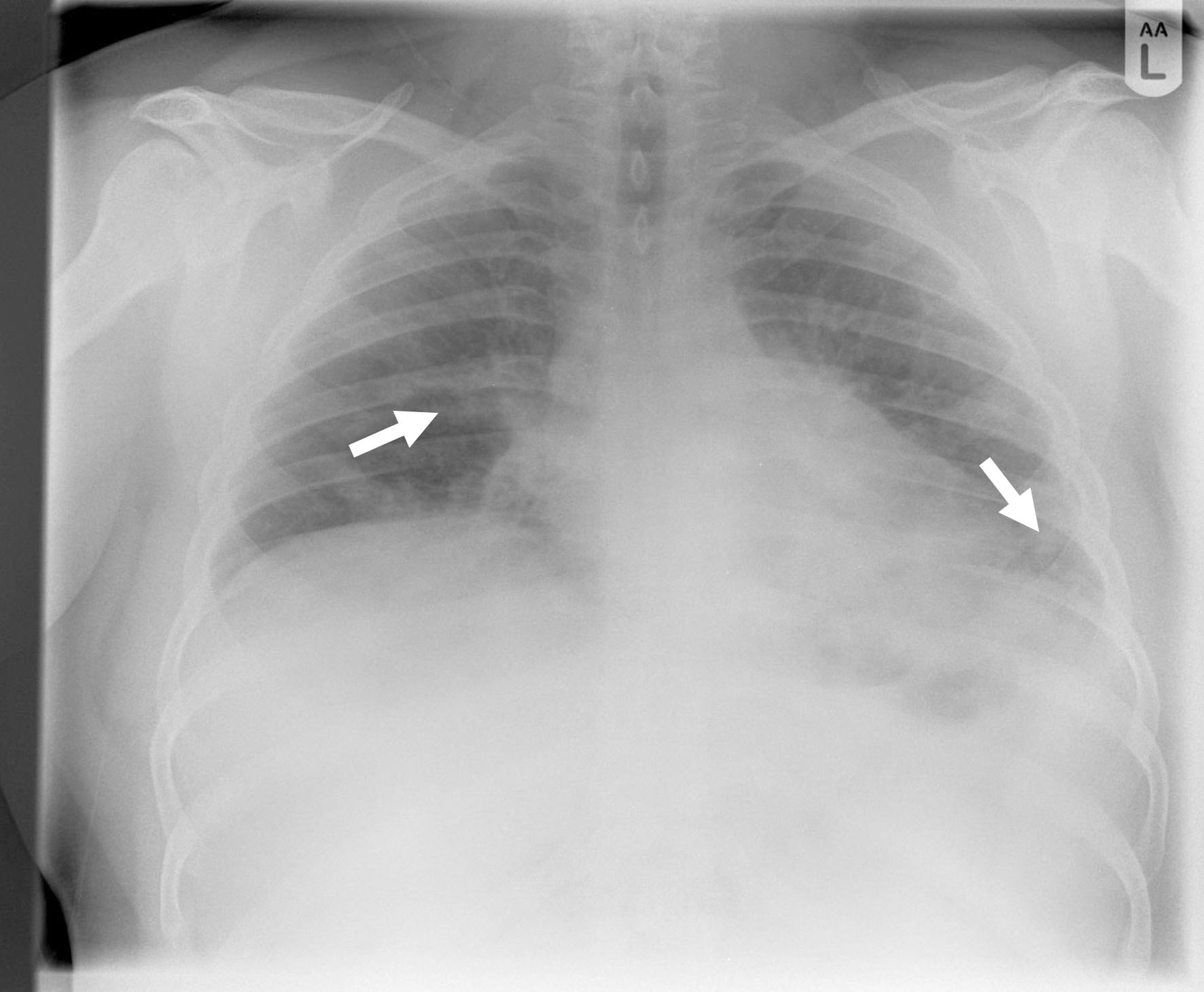
Figure 1. Chest X-ray showing patchy air space shadowing in both lungs, prominently in mid and lower zones (arrows).
Figure 2.
Figure 2. Computed tomography pulmonary angiography showing no pulmonary embolism but multiple ground glass lesions covering more than 50% of the alveolar surface, concerning for pulmonary infection including moderate form of covid pneumonitis (arrows).
Figure 3.
Figure 3. First follow-up computed tomography chest showing improvement of ground glass appearance (arrows).
Figure 4.
Figure 4. Diffuse patchy lung infiltrates, predominantly in both lung bases, with some variation from previous imaging (arrows).
Figure 5.
Figure 5. Magnetic resonance imaging of femur with evidence of myositis (arrows).